Richard Conniff's Blog, page 73
May 24, 2013
Introducing Antibiotic Resistance to the Wild

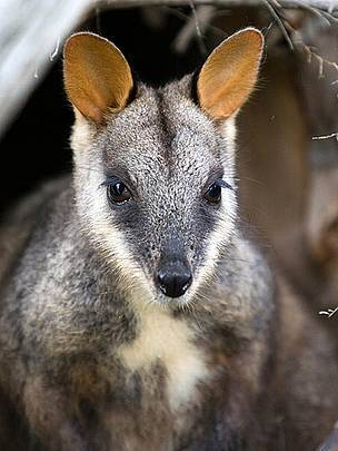
Cute but a carrier (Photo: Ben Bishop/WWF-Aus.)
A few weeks ago, I reported that antibiotic resistance had jumped from humans to wildlife in a remote national park in southern Africa. Now a new paper reports that it is happening in Australia, too, by way of a captive-breeding reintroduction program.
Here’s the story from PLOS ONE, where the study is being published:
Endangered brush-tail rock wallabies raised in captive breeding programs carry antibiotic resistance genes in their gut bacteria and may be able to transmit these genes into wild populations, according to research published May 22 in the open access journal PLOS ONE by Michelle Power and colleagues from Macquarie University in New South Wales, Australia.
Brush-tail rock wallabies are currently being raised in species recovery programs and restored to the wild to bolster populations of this endangered species. Here, researchers found that nearly half of fecal samples from wallabies raised in these programs contained bacterial genes that encode resistance to streptomycin, spectinomycin and trimethoprim. None of these genes were detected in samples from five wild populations of wallabies. The authors add, “How these genes made their way into the wallaby microbes is unknown, but it seems likely that water or feed may have acted as a conduit for bacteria carrying these genes.”
Previous research shows that proximity to humans can increase animals’ exposure to antibiotic resistance genes and the organisms that carry them. Antibiotic resistant bacteria have been reported in the wild from chimpanzees in Uganda, Atlantic bottlenose dolphins and a wide range of fish, birds and mammals. According to the researchers, their findings highlight the potential for genes and pathogens from human sources to be spread. Power says, “We found that antibiotic resistance genes from human pathogens have been picked up by endangered rock wallabies in a breeding program, and may spread into the wild when the wallabies are released.”
Source: Michelle L. Power, Samantha Emery, Michael R. Gillings. Into the Wild: Dissemination of Antibiotic Resistance Determinants via a Species Recovery Program. PLoS ONE, 2013; 8 (5): e63017 DOI: 10.1371/journal.pone.0063017


May 23, 2013
The Wildlife Microbiome: A New Way of Understanding Species
Here’s my latest piece for Yale Environment 360:
A few years ago, as he was puzzling over the egg-tending behavior of a common forest salamander, herpetologist Reid Harris began to wonder if he might be looking at a novel solution to one of the most devastating global pandemics of our time. No one knew it at the time, but the four-toed salamander was about to become a pioneer species in the incipient field of microbial conservation biology, a dramatically different way of understanding and protecting wildlife.
The salamander Harris was studying lives in the leaf litter around forest pools from Michigan to Florida. Not all females in the species tend their nests, but the ones that do have an oddball way of weaving in and out among the eggs.
In 2008, Harris and other researchers at James Madison University in Virginia demonstrated that this weaving behavior serves to inoculate the eggs with antifungal bacteria from the female’s skin — and that the ones receiving this form of probiotic maternal care have a much higher rate of survival against infection by a common egg fungus.
At a herpetological conference three years earlier, Harris had suggested that it might be possible to identify anti-fungal bacteria species occurring naturally on the skin of other amphibian species and “bio-augment” them as a probiotic protection. And he thought it might work not just against the egg fungus, but also against the chytrid fungus that is causing massive declines in amphibian species worldwide. That fungus has rapidly spread around the world over the past two decades and now affects more than 500 amphibian species in 52 countries. Spores from the chytrid fungus, Batrachochytrium dendrobatidis (or Bd), invade the skin of amphibians and block normal respiration, leading to electrolyte imbalance, brain swelling, and death.
The chytrid fungus has already pushed at least two species over the edge into extinction (the golden toad in the Costa Rican cloud forest, and the gastric brooding frog in Australia). Though scientists are cautious about saying such things categorically, the pandemic may have caused as many as 100 extinctions, with gloomy herpetologists predicting many more still to come.
In any less desperate group of biologists, what Harris was proposing would probably have elicited a faint rolling of the eyes. Asked about the idea of using microbes to protect animals in the field and at zoos, for instance, one zoo curator interviewed for this article remarked that conservationists already have plenty to worry about “without taking some kind of biochemistry approach.”
But just within the past decade, rapid improvements in DNA sequencing technology have made it possible and economically practical to identify every kind of bacteria, fungi, and virus living in and around a species. (In the past, biologists could study only the small percentage of microbes that can be cultured in a Petri dish.) Researchers have so far applied this new technology mainly to the human microbial community, or microbiome, and that research has already produced a dramatic shift in thinking about our own health care. In place of the old germ theory view of microbes as our deadly enemy, it now looks as if they are also our essential allies, both a cause of diseases and a key to preventing them.
But “the professional conservation community appears to be largely unaware of these developments,” a team of biologists noted last year in the journal Conservation Biology. The article proposed “that the concepts and methods used to study the human microbiome could be applied to meet conservation challenges” — for instance, to understand why the same Helicobacter bacteria that are harmless in the wild cause gastritis for cheetahs in zoos, or to figure out how to prevent the callithricid wasting syndrome that afflicts captive marmosets and tamarins.
Harris’s idea about amphibian species caused a light to switch on for Vance Vredenburg of San Francisco State University. He soon struck up a partnership with Harris to test the probiotic idea in the mountains of the Sierra Nevada. There, Vredenburg had been watching helplessly as the chytrid fungus swept through, killing off population after population of mountain yellow-legged frogs.
The skins of yellow-leg frogs turned out to carry the same anti-fungal bacteria species that Harris had found on salamanders back East, though not necessarily enough to protect them. The researchers began to brew the stuff in the lab, producing buckets of murky purple bacterial soup. (Janthinobacterium lividum gets its species name from its bluish color.) Then they briefly bathed lab-reared yellow-legged frogs in the liquid and, after waiting two days for the probiotic to build up on the skin, they exposed the frogs to the chytrid fungus. In the group that had not gotten the protective bath, more than 80 percent soon died. In the group treated with anti-fungal bacteria, none did.
Vredenburg was already thinking about field-testing microbial protection on a pond at 11,000 feet in Sequoia & Kings Canyon National Parks, one of the last remaining havens for the frogs, where the chytrid fungus was soon expected to arrive.
The idea of conservation biologists turning to microbes in the fight against a pandemic probably should not sound all that surprising. Much of the recent reporting about the microbiome in the popular press has made it sound as if this is a bold new world discovered by the medical community. But microbiome research actually originated in the ecological community, says Yale University microbiologist Jo Handelsman. Ecologists started thinking about the beneficial role of microbes at least 130 years ago, she says, when they identified the crucial function of bacteria in fixing nitrogen for soybeans, peanuts, and other legumes. It’s the medical community that has borrowed ecological thinking, she argues, not the other way around.
Even so, the idea of microbial conservation biology may seem like a leap for wildlife scientists because it requires them to think about their familiar study animals not just as organisms, but as superorganisms — that is, as the host species together with its essential bacteria, viruses, and fungi. It requires thinking not just about the genome but about the metagenome, a term Handelsman coined for the interacting genes of the host species and all its microbial fellow travelers.
It’s too soon to say exactly how this new way of looking at species will change how we treat wildlife. But for instance, understanding the microbiome could help improve the generally dismal success rate of efforts to reintroduce captive-bred species to the wild, biologist Kent H. Redford, lead author of the Conservation Biology article, suggested in an interview. Maybe “the zoo zebra you put back in the wild isn’t really a zebra,” he said, because it lacks certain crucial microbes from its native habitat.
For instance, hellbenders, a kind of giant salamander, have rapidly declined in streams from New York and Ohio down to Mississippi. They are now being captive-bred in zoos for reintroduction. But their young “are likely to have atypical and depauperate microbial communities and naive immune defences,” according to another article, being published in the current issue of Ecology Letters. Probiotic inoculation might help them handle the stress of being released into the wild, writes lead author Molly C. Bletz, a researcher in Harris’s lab at James Madison University.
Though the idea has not yet been tested, Bletz and her co-authors also suggest that probiotic inoculation might provide protection against white-nose syndrome, which has devastated bat populations in the eastern United States. They recommend screening European bats to see if some kind of protective microbe might be part of the explanation for why bats there survive infection with the white-nose pathogen, while bats in North America succumb.
Other researchers are also working on the microbiome in various wildlife species simply to establish a microbial baseline, or to get a microbial perspective on a particular biological process. At the University of Colorado
in Boulder, for instance, researchers recently compared the noxious oral bacteria of Komodo dragons in the wild with the somewhat less noxious bacteria of their captive counterparts at the Denver Zoo. Another study in the same lab looked at the role of microbes in the feast-and-famine eating cycle of Burmese pythons. (To be sure that they were looking at a python’s own microbes, says researcher Liz Costello, they first had to characterize the microbes in the food it was eating. That meant putting frozen white rats in a blender and liquefying them for analysis. “It looked like a chocolate milkshake, with teeth in it.”)
And in a recent issue of the journal Science, Michigan State University researchers reported that they have developed a technique for preventing a mosquito species from acquiring and transmitting malaria by inoculating it with a particular strain of Wolbachia bacteria. The same group successfully field-tested a similar technique in 2011 to prevent transmission of dengue fever.
For Harris’s probiotic idea, the only field test so far is the one the National Park Service gave Vredenburg permission to try in 2010 in California, at Sequoia & Kings Canyon parks. As a precaution against introducing an unfamiliar microbe, he first visited Dusy Basin there that July and found the right bacteria on just one of 40 frogs. Over the next ten days, he brewed the bacteria from a single droplet into tens of billions of bacterial cells, about 10 liters of liquid. Then he returned to inoculate 80 percent of the frog population, leaving the rest untreated as a control. When the chytrid fungus arrived as expected later that summer, only the treated frogs survived.
For Vredenburg, killing weather the following winter and predation by introduced species took some of the satisfaction off that initial success. Only a few yellow-legged frogs have managed to hang on at Dusy Basin. And when another researcher subsequently attempted to inoculate laboratory-reared Panamanian golden frogs against the chytrid fungus using the same bacterial species, J. lividum, the experiment failed completely. To Harris, that merely suggests the need to do more careful preliminary research to identify the right bacteria for a particular species and habitat.


Coal: The Biodiversity Fuel
Paradise for a strange new world.
Before we get started, a warning. What you’re about to read is going to sound at first like something cooked up by the same folks who gave us the oxymoronic (and otherwise moronic) advertising slogan “Clean Coal.” It will sound like a fantasy story even a Fox News anchor would not dare announce: “Coal—The Biodiversity Fuel.”
In a paper being published in the journal Biological Conservation, researchers in the Czech Republic, who have been studying bees and wasps, report that some of that country’s endangered species, including four insects that had been presumed regionally extinct, have turned up instead thriving in the fly ash heaps at coal-fired power plants.
Fly ash, as the paper helpfully explains, is what’s left over after a power plant burns coal, and it’s composed of “glass-like particles of mineral residua which are carried out of the boiler in the flow of exhaust gases,” plus bottom ash, boiler slag, and “flue gas desulphurization materials.” To be clear, the “fly” in “fly ash” is not a reference to insects; rather, it has to do with the fact that the substance is so light and fine that it flies up during combustion.
The study found 227 species of bees and wasps, including 35 that were endangered or critically endangered, living at two power plant sites. Some of these insects are important pollinators, and others may be valuable as predators and parasitoids for controlling agricultural pests.
To read the rest of this story click here.


May 15, 2013
Grandma’s Pregnancy Test and the Extinction of Species

An African clawed frog, the species that introduced the chytrid fungus to the world. (Photo: H. Krisp/Wikimedia Commons)
Ever since The Hot Zone became a non-fiction bestseller 20 years ago, people have been fretting about the likelihood that an emerging pathogen could cause a global pandemic, killing tens of millions of humans. But they seldom pause to consider that it is already happening in the animal world—or that the pandemic that’s now decimating frogs and other amphibians provides a perfect model for how it could also happen to us.
The pathogen this time is the chytrid fungus, which has raced around the world over the past two decades, and now afflicts more than 500 amphibian species in 52 countries. When spores of this fungus penetrate a victim’s skin, a slough of dead cells builds up on the surface, blocking respiration. The electrolytes go out of balance. The brain swells. The frog sits with its legs skewed out oddly to the sides. Death soon follows, often for an entire community of amphibians around a pond or wetland. The chorus of peepers goes silent.
“I can’t think of another disease on the planet more significant than this amphibian disease,” says Peter Daszak, president of EcoHealth Alliance, a New York-based group focused on the role of the wildlife trade in the introduction of dangerous pathogens. “No disease of humans has ever wiped us out.” But he estimates that the chytrid fungus pandemic has already caused the extinction of more than 100 species, including the golden toad in the cloud forests of Costa Rica, the gastric brooding frog in Queensland, Australia, and 20 or 30 species of brilliantly colored Harlequin frogs in Central and South America. “And it’s still causing extinctions.”
A paper being published today in the science journal PLOS ONE adds new evidence to the story of how innocent and even seemingly humane missteps unleashed this killer on the amphibian world. Maybe it’s best to start with our own families: For anyone born from the 1920s up until the late 1970s, the way our mothers generally got the happy news of our impending arrival was by way of the African clawed frog, an East African species in the genus Xenopus. … click here to read the rest of the story.


May 10, 2013
Madness in the Village of Elephants

Andrea Turkalo at the observation post that has now become a firing line.
In the forest clearing locals call the “Village of Elephants,” or Dzanga Bai, 17 heavily armed men arrived this past Wednesday, May 8, with AK-47s. They were bound for the observation tower where tourists in the Central African Republic have often come to admire the forest elephants, and where researchers have worked to decipher the language of elephants for more than 20 years.
It was over in a few horrific minutes.
When guards who had previously been disarmed by rebel forces went back yesterday, May 9, they counted the butchered carcasses of 26 elephants killed for their ivory, including four babies.
The killing happened in the Dzanga-Sangha Protected Area, in the southwest corner of the country, on the border with Cameroon and the Democratic Republic of the Congo. The Dzanga Bai itself is a UNESCO World Heritage Site. In 2010, the CBS show 60 Minutes described it as “one of the most magical places on Earth.”
At least for the moment, … to read the full story click here.


May 9, 2013
Jurassic Park and the Fear of Feathers

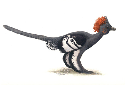
Anchiornis huxleyi in full feather (Illustration: Michael DiGiorgio)
The much-delayed Jurassic Park 4 sequel was delayed again early this week, and it’s tempting to imagine that animal science might be the reason. (Okay, tempting and really, really stupid, but indulge me for a bit.) This lucrative movie franchise dates back 20 years now, to 1993, which is like saying it started somewhere in the Cenozoic as far as our understanding of dinosaurs goes.
When the original Jurassic Park was still in its first theatrical run, paleontologists were already digging up what has since become a gaudy parade of fossils demonstrating that dinosaurs were in fact frequently tricked out with feathers, feather-like filaments, and even a three-inch-thick coating of “dino fuzz.”
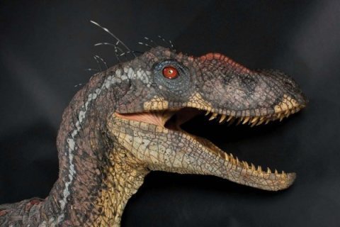
Universal Pictures grudgingly acknowledged this new science when it released Jurassic Park III in 2001. Like an anxious parent in the Punk Rock era, it allowed Velociraptor to flaunt a miserable little mohawk of about a dozen filaments sprouting out of the top of its head.
But otherwise the franchise has conformed to the stereotype of dinosaurs as scaly, naked red-eyed monsters. And for an obvious reason: A Tyrannosaurus rex that looked like Big Bird might not have audiences wetting their pants in the balcony, or opening their wallets at the box office. So back in March, Colin Trevorrow, tapped as the latest director in the series, tweeted: “No feathers. #JP4”
But maybe now he’s gone back for a re-think.

A Hollywood velociraptor
Here’s where the fossil evidence currently stands, as outlined by Julia Clarke, a University of Texas paleontologist who is also the author of an article “Feathers Before Flight,” appearing today, May 9, in the journal Science: Paleontologists have been thinking about the connection between dinosaurs and living birds since the discovery of the fossil bird Archaeopteryx back in 1861, and especially since 1970, when John Ostrom at Yale University pointed out the many similarities between bird skeletons and those of the Theropoda dinosaurs, including T. rex and Velociraptor.
The current revolution began in the mid-1990s, when … to read the full article click here.


May 8, 2013
The Body Eclectic on C-SPAN’s Washington Journal
http://www.c-span.org/Journal/
On “Washington Journal” this morning, Greta Brawner interviewed me about my microbiome story in the May issue of Smithsonian. You can see the show here. You should be able to scroll down on the right to get to my segment. Look carefully and see me not smile for 42 minutes!


May 7, 2013
Shape Shifter

(Photo: Cally Harper)
Tongues can do delightful and astonishing things. I am thinking of the way a frog fires its sticky tongue halfway across the universe to snag a passing insect (see below). Or how an alligator snapping turtle wriggles its tongue like a worm as a dinner invitation to fish. And now the Pallas’s long-tongued bat (Glossophaga sorcina) joins this elite club of astonishing animal tongue artists.
These bats, found from Argentina to northern Mexico, and sometimes into Arizona and New Mexico, have the fastest metabolism ever recorded in a mammal, says a 2007 study in the journal Nature. They burn half their body fat each day, and have to make up for it at night by consuming as much as 150 percent of their body weight in nectar from flowers. And of course, they have to do it on the wing. According to a study published today in the Proceedings of the National Academy of Science, the secret to its success is a remarkable ability to change the shape of its tongue into a hemodynamic—or blood-swollen—“nectar mop.”
When lead author Cally Harper, a doctoral candidate in biomechanics at Brown University in Rhode Island, began her study, specialists already knew that bats of this species have an unusual fringe of hair-like structures around the tip of the tongue. They assumed these were useful for collecting nectar—but passively, like raking icing off a cake using your fingernails. Biologists also knew that these bats have enlarged blood vessels in their tongues. But they didn’t know what to make of them. Harper had a hunch that the two features might be connected, especially since … Read the full article here.


A Forgotten Pioneer of Vaccines

Jeryl Lynn Hilleman with her sister, Kirsten, in 1966 as a doctor gave her the mumps vaccine developed by their father.
This is a piece I wrote for today’s New York Times. It briefly appeared in the the front page online, then the world righted itself, and it gave way to an item about Ru Paul, and then one about Madonna.
We live in an epidemiological bubble and are for the most part blissfully unaware of it. Diseases that were routine hazards of childhood for many Americans living today now seem like ancient history. And where every mother could once identify measles in a heartbeat, even the best hospitals have to call in their eldest staffers to ask: “Is this what we think it is?”
To a remarkable extent, we owe our well-being, and in many cases our lives, to the work of one man and to events that happened 50 years ago this spring.
At 1 a.m. on March 21, 1963, an intense, irascible but modest Merck scientist named Maurice R. Hilleman was asleep at his home in the Philadelphia suburb of Lafayette Hill when his 5-year-old daughter, Jeryl Lynn, woke him with a sore throat. Dr. Hilleman felt the side of her face and then the telltale lump beneath the jaw. He tucked her back into bed, about the only treatment anyone could offer at the time.
For most children, mumps was a nuisance disease, nothing worse than a painful swelling of the salivary glands. But Dr. Hilleman knew it could sometimes leave a child deaf or otherwise permanently impaired.
He alerted the housekeeper that he was going out (his wife had recently died of breast cancer) and then drove 20 minutes to pick up proper sampling equipment from his laboratory. Back at the house, he woke Jeryl Lynn long enough to swab the back of her throat and immerse the specimen in a nutrient broth. Then he drove back to store it in the laboratory freezer.
The name Maurice Hilleman may not ring a bell. But today 95 percent of American children receive the M.M.R. — the vaccine for measles, mumps and rubella that Dr. Hilleman invented, starting with the mumps strain he collected that night from his daughter.
It was by no means his only contribution. At Dr. Hilleman’s death in 2005, other researchers credited him with having saved more lives than any other scientist in the 20th century. Over his career, he devised or substantially improved more than 25 vaccines, including 9 of the 14 now routinely recommended for children.
“One person did that!” said Dr. Anthony S. Fauci, a longtime friend of Dr. Hilleman’s and now director of the National Institute of Allergy and Infectious Diseases. “Truly amazing.”
As a young man in Montana, Maurice Hilleman had intended only to become a manager at the J..C. Penney store. He turned out not to have the perfect retail personality. (Asked later in life what he was proudest of in his career, he replied, “Being able to survive while being a bastard.”)
He went on to spend most of his career at Merck, but the corporate personality also eluded him. He had a sailor’s vocabulary, and his brand of peer review often included the phrase“You’re full of crap” (though he said it “in a constructive way,” Dr. Fauci said with a smile).
But everyone recognized Dr. Hilleman’s genius at discovering and perfecting vaccines, which he pursued, Dr. Fauci said, with a rare combination of “exquisite scientific knowledge” and “amazingly practical, get-it-done personality.”
A vaccine is a tool for coaxing the immune system to resist a disease without having to experience the actual symptoms, and making them was an art as much as a science. “It’s not like there was a formula for this,” said Dr. Paul Offit, a Philadelphia pediatrician, vaccine developer and author of “Vaccinated,” a 2007 biography of Dr. Hilleman.
The general practice was to isolate a disease organism, figure out how to keep it alive in the laboratory, then weaken or “attenuate” it by passing it over and over through a series of cells, typically from chicken embryos, until it could no longer reproduce in humans, but could still elicit an immune response. Other steps followed, particularly for Dr. Hilleman, who was obsessed with safety and with stripping away unwanted side effects.
That spring of 1963, the Food and Drug Administsation also granted the first license for a vaccine against measles. Much of the early work on the virus had been done in the laboratory of John Enders at Boston Children’s Hospital, but the vaccine still commonly produced rashes and fevers when Dr. Hilleman began to work on it.
Under pressure from public health officials to stop a disease then killing more than 500 American children every year, Dr. Hilleman and Dr. Joseph Stokes, a pediatrician, devised a way to minimize the side effects by giving a gamma globulin shot in one arm and the measles vaccine in the other. It was the beginning of the end of the disease in this country.
Dr. Hilleman then went on to refine the vaccine over the next four years, eventually producing the much safer Moraten strain, which is still in use today. As always, he kept himself in the background: The name stands for “More Attenuated Enders.”
One other crucial event in the development of M.M.R. happened that spring of 1963: An epidemic of rubella began in Europe and quickly swept around the globe. In this country, the devastating effect on first- trimester pregnancies caused about 11,000 newborns to die, according to the U.S. Centers for Disease Control and Prevention. Another 20,000 suffered birth defects, including deafness, heart disease, and cataracts.
Dr. Hilleman was already testing his own vaccine as the epidemic ended in 1965. But he agreed to work instead with a vaccine being developed by federal regulators. It was, he later recalled, “toxic, toxic, toxic.” By 1969, he had cleaned it up enough to obtain a license and prevent another rubella epidemic. Finally, in 1971, he put his measles, mumps, and rubella vaccines together to make M.M.R., replacing a series of six shots with just two.
Or rather not finally. In 1978, having found a better rubella vaccine than his own, Dr. Hilleman phoned its developer to ask if he could use it in the M.M.R. The developer, Dr. Stanley Plotkin, then of the Wistar Institute in Philadelphia, was momentarily speechless. It was an expensive choice for Merck, and might have been a painful one for anyone other than Dr. Hilleman.
“It’s not that he didn’t have an ego. He certainly did,” Dr. Plotkin recalled in a recent interview. “But he valued excellence above that. Once he decided that this strain was better, he did what he had to do,” even if it meant sacrificing his own work.
Given Dr. Hilleman’s obsession with safety and effectiveness, it came as a bitter surprise toward the end of his life when his M.M.R. became the center of what Dr. Offit called “a perfect storm of fear” about vaccines. It arose partly because the very success of vaccines had made people forget just how devastating the diseases they prevented could be. “You’re asking parents to protect their children from diseases most people no longer see,” says Dr. Offit, “using a biological fluid that most parents don’t understand.”
Then, on top of this collective forgetting, in 1998 The Lancet, a respected British medical journal, published an article alleging that the M.M.R. had caused an epidemic of autism.
The lead author, Dr. Andrew Wakefield, became a media celebrity, and ostensibly educated parents began to balk at having their children immunized. Dr. Hilleman, who might reasonably have been expected to win a Nobel Prize, got hate mail and death threats instead.
Multiple independent studies would eventually demonstrate that there is no link between M.M.R. and autism, and Dr. Wakefield’s work has been widely discredited. In 2010, the British medical authorities stripped him of the right to practice medicine, and The Lancet belatedly retracted the 1998 article.
It came too late, not just for Dr. Hilleman, who by then had died of cancer, but also for many parents who mistakenly believed that avoiding the vaccine was the right way to protect their children. In 2011 alone, a measles outbreak in Europe sickened 26,000 people and killed 9. . Because the disease is contagious enough to pick up from a traveler walking by in the airport, cases still also occur in this country among the unvaccinated.
But Dr. Hilleman would probably still find reason to be encouraged. The Measles and Rubella Initiative, a global campaign organized in 2001, has given the M.M.R. vaccine to a billion children in this century, preventing 9.6 million deaths from measles alone, for less than $2 a dose. According to Dr. Stephen L. Cochi, a global immunization adviser at the C.D.C., the initiative is “on the verge of setting a target date” to eradicate the disease.
In this country, the strain that Dr. Hilleman collected from his daughter that night in 1963 has reduced the incidence of mumps to fewer than 1,000 cases a year, from 186,000. Characteristically, he named it not for himself but for his daughter. And Jeryl Lynn Hilleman, now a financial consultant to biotech startups in Silicon Valley, turns the credit back on her father.
He was driven, she said in an interview, “by a need to be of use — of use to people, of use to humanity.”
“All I did,” she added, “was get sick at the right time, with the right virus, with the right father.”


May 2, 2013
Attack of the Killer Shrimp

Come closer, see how pretty I am. (Photo: Michael Bok)
This is my latest post for TakePart:
One day early this year, on the Connecticut beach where I have walked most days for the past 15 years, I came across an animal I’d never seen before, washed up in the seaweed. At first, a neighbor and I thought it might be an immature lobster. It was about eight inches long, with a greenish-gray segmented carapace, and goggle eyes mounted on stalks. But in place of a lobster’s formidable claws, it seemed to have only a couple of feathery antennae.
So: Lobster-like, but lame.
Lord, were we ever wrong. It was in fact one of the most violent creatures on Earth, “enchantingly violent,” in the words of a biologist who studies them, violent enough to bring to mind the old “Jaws” soundtrack (DUNT-dunt, DUNT-dunt) and the teaser line: “You’ll never go in the water again!”
It was a mantis shrimp, so named because many people think they look like a cross between a preying mantis and a shrimp, though they are actually members of their own crustacean order, the Stomapoda. There are about 400 species of mantis shrimp and they inhabit coastlines worldwide, leading mostly solitary lives, typically burrowing in mud and silt on the sea floor, or hiding out in rocky formations.
But let’s get to the violence. Click hear to read the rest of the story.


