
Jeffrey Dach's Blog, page 4
December 3, 2024
Are Spider Silk Gene Sequences in the C0\/lD \/axxine Causing Fibrous Blood Clots?
 Are Spider Silk Gene Sequences in the
C0\/lD \/axxine
Causing Fibrous Blood Clots?
by Jeffrey Dach MD
Sarah, a long term patient in my office doing well on bioidentical hormone replacement told me a story about her cousin who developed blood clots after a C0\/lD booster, requiring abdominal surgery for mesenteric artery occlusion. Thankfully, her cousin is back home recuperating. Since the rollout of the
C0\/lD \/a<
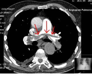
Header Image: IV contrast enhanced CAT scan showing Saddle Embolus (Red Arrows blood clot) lodged across bifurcation of pulmonary artery, Transferred from en.wikipedia to Commons. Author Glitzy queen00 at English Wikipedia. public domain. wikimedia commons.
Watch video of Richard Hirschman, Funeral Director And Embalmer Removing Clot From Jugular Vein:
Pathologist Ryan Cole MD, Discusses Unusual Blood Clots which contain amyloid protein: Link
Pathologist, Dr. Ryan Cole, tells Dr. Drew about the strange, foot-long cl0ts he’s been finding in mRNA injected patients—both alive and deceased—and how they can be broken down and cleared from the body using a natural enzyme called ‘Nattokinase’.
“The morticians that started seeing these—when a body comes in and they have to preserve it, they cannulate large vessels, they put their needles in large vessels—they started getting back pressure that they hadn’t experienced before. And there are one or two that have spoken out, but I know of about another 50 that are seeing the same, who want to keep their jobs, so they don’t say anything.”
Biologist Prof. Dr. Ulrike Kammerer, University of Würzburg
Watch this video in which Dr. Ulrike Kammerer says C0\/lD jabs contain spider silk gene sequence, the genetic code for spider silk production (Spidroins which stain for amyloid). Spider silk protein has been the subject of extensive research for the last twenty years and has been successfully produced using recombinant genetic engineering techniques. So, inserting the gene sequence for spider protein is a simple “off the shelf” maneuver to weaponize the C0\/lD \/a<
DNA Contamination of mRNA C0\/lD \/a<
A new study from Germany by Dr. Ulrike Kämmerer published in Science, Public Health Policy and the Law December 3, 2024 found high levels of residual DNA in the mRNA
C0\/lD \/a<
Decades of gene therapy research has shown DNA contamination causes insertional mutagenesis, and cancer. Dr. Ulrike Kämmerer’s study calls for halting the use of mRNA C0\/lD \/a<guidance, September 13, 2023, recommending against use of mRNA C0\/lD \/a<
Conclusion: Spider silk protein has been the subject of extensive research for the last twenty years and has been successfully produced using recombinant genetic engineering techniques. So, inserting the gene sequence for spider protein is a simple “off the shelf” maneuver to weaponize the C0\/lD \/a<
Articles with Related interest:
Florida Department of Health Advises Against C0\/lD B00STERS
Dr. Karina Whitehouse Presentation on mRNA \/axxines
Jeffrey Dach MD
7450 Griffin Road, Suite 190
Davie, Fl 33314
954-792-4663
References
1) C0\/lD Exclusive: 70% of Embalmers Report Finding Strange Blood Clots Beginning in Mid-2021, Childrens Health Defense by John-Michael Dumais
January 22, 2024
2) Embalmers are Continuing to Find Mysterious Clots in the Vaccinated...Reviewing the results of a recent citizen’s investigation and what we now know about these amyloid clots.
A Midwestern Doctor Feb 22, 2024
3) BioNTech RNA-Based COVID-19 Injections Contain Large Amounts Of Residual DNA Including An SV40 Promoter/Enhancer Sequence
Ulrike Kämmerer Verena Schulz Klaus Steger * Peer Reviewed, Clinical Research
Science, Public Health Policy and the Law 12/03/2024
The study calls for halting the use of mRNA vaccines until these safety issues are thoroughly investigated and resolved.
Conclusion of the study: The findings raise significant safety concerns about the BNT162b2 vaccine due to high levels of residual DNA found in vials, which far exceeds safety limits. The study calls for halting the use of mRNA vaccines until these safety issues are thoroughly investigated and resolved.
4) Just In: Peer-Reviewed German Study Reveals mRNA Vaccine DNA Contamination Exceeds Safety Limits; German Scientists Urge IMMEDIATE HALT!
We call for an immediate halt of all RNA-based biologicals until these concerns are scientifically addressed and convincingly dispelled.
Aussie17 Dec 03, 2024
5) BREAKING: German Study Raises Grave Safety Concerns over BioNTech’s Comirnaty Sonia Elijah Dec 03, 2024
6) Arndt, Tina, et al. “Spidroin N-terminal domain forms amyloid-like fibril based hydrogels and provides a protein immobilization platform.” Nature Communications 13.1 (2022): 4695.
7) Qi, Xingmei, et al. “Spider Silk Protein Forms Amyloid‐Like Nanofibrils through a Non‐Nucleation‐Dependent Polymerization Mechanism.” Small 19.46 (2023): 2304031.
8) Kenney, John M., et al. “Amyloidogenic nature of spider silk.” European journal of biochemistry 269.16 (2002): 4159-4163.
9) Dai, Bin, et al. “Fibril self-assembly of amyloid–spider silk block polypeptides.” Biomacromolecules 20.5 (2019): 2015-2023.
10) Abelein, Axel, et al. “High-yield production of amyloid-β peptide enabled by a customized spider silk domain.” Scientific reports 10.1 (2020): 235.
11) Rising, Anna, et al. “Spider silk proteins: recent advances in recombinant production, structure–function relationships and biomedical applications.” Cellular and Molecular Life Sciences 68 (2011): 169-184.
12)
Spider Silk Gene Sequence Found In COVID Jabs April 21, 2024
13) Biologist Prof. Dr. Ulrike Kammerer: C0\/lD jabs contain spider silk gene sequence
————————–
14) Spider silk gene sequence found in COVID jabs. GMO spider silk goats, bacteria, worms already exist
15) Scheibel, Thomas. “Spider silks: recombinant synthesis, assembly, spinning, and engineering of synthetic proteins.” Microbial cell factories 3 (2004): 1-10.
16) Abelein, Axel, et al. “High-yield production of amyloid-β peptide enabled by a customized spider silk domain.” Scientific reports 10.1 (2020): 235.
17) Kenney, John M., et al. “Amyloidogenic nature of spider silk.” European journal of biochemistry 269.16 (2002): 4159-4163.
18) Scheibel, Thomas. “Spider silks: recombinant synthesis, assembly, spinning, and engineering of synthetic proteins.” Microbial cell factories 3 (2004): 1-10.
19) Qi, Xingmei, et al. “Spider silk protein forms amyloid‐like nanofibrils through a non‐nucleation‐dependent polymerization mechanism.” Small 19.46 (2023): 2304031.
20) Tjernberg, L. O., et al. “Transmissible amyloid.” Journal of Internal Medicine 280.2 (2016): 153-163.
spidroins are highly soluble and form disordered and partly α-helical structures, but upon passage through the spinning duct, they are converted into solid fibres made up of amyloid-like β-sheets and amorphous parts 48-51. Thus, like amyloid fibrils, silk fibres are formed from soluble proteins under physiological conditions 52, but there are also marked differences between amyloid and silk. Amyloid fibrils are several orders of magnitude smaller and their β-strands are oriented perpendicular to the fibre axis, unlike spider and silkworm silk in which the direction of the β-strands is parallel to the fibre axis 50, 53-55. Furthermore, in contrast to silk proteins, amyloid-forming proteins in their natively folded state generally have specific functions unrelated to fibre formation 56.
21) Kong, Na. “General Methods to Produce and Assemble Recombinant Spider Silk Proteins.” Fibrous Proteins: Design, Synthesis, and Assembly (2021): 57-67.
22) Roth, David Eugene. Genetic Engineering of Functional Large Amyloid Fibers. Diss. Virginia Tech, 2016.
The experimental results show that large amyloid fibers with predictable size and mechanical properties can be anticipated and encoded at the genetic level
23) Pretorius, Etheresia, et al. “Prevalence of amyloid blood clots in COVID-19 plasma.” medrxiv (2020): 2020-07.
We show here that microclots can be detected in the native plasma of COVID-19 patient, and in particular that such clots are amyloid in nature as judged by a standard fluorogenic stain. This provides a rapid and convenient test (P<0.0001), and suggests that the early detection and prevention of such clotting could have an important role in therapy.
24) Exclusive: 70 percent of embalmers report finding strange blood clots beginning in mid-2021
25) Acevedo-Whitehouse, K., and R. Bruno. “Potential health risks of mRNA-based vaccine therapy: a hypothesis.” Medical Hypotheses 171 (2023): 111015.
We propose that in susceptible individuals, cytosolic clearance of nucleotide modified synthetic (nms-mRNAs) is impeded. Sustained presence of nms-mRNA in the cytoplasm deregulates and activates endogenous transposable elements (TEs), causing some of the mRNA copies to be reverse transcribed. The cytosolic accumulation of the nms-mRNA and the reverse transcribed cDNA molecules activates RNA and DNA sensory pathways. Their concurrent activation initiates a synchronized innate response against non-self nucleic acids, prompting type-I interferon and pro-inflammatory cytokine production which, if unregulated, leads to autoinflammatory and autoimmune conditions, while activated TEs increase the risk of insertional mutagenesis of the reverse transcribed molecules, which can disrupt coding regions, enhance the risk of mutations in tumour suppressor genes, and lead to sustained DNA damage. Susceptible individuals would then expectedly have an increased risk of DNA damage, chronic autoinflammation, autoimmunity and cancer. In light of the current mass administration of nms-mRNA vaccines, it is essential and urgent to fully understand the intracellular cascades initiated by cellular uptake of synthetic mRNA and the consequences of these molecular events.
26) Mead, M. Natnaniel, et al. “COVID-19 Modified mRNA “Vaccines” Part 1: Lessons Learned from Clinical Trials, Mass Vaccination, and Censorship by the Bio-Pharmaceutical Complex.” International Journal of Vaccine Theory, Practice, and Research 3.1 (2023): 1112-1178.
27) Mead, M. Nathaniel, et al. “COVID-19 modified mRNA “vaccines”: Lessons learned from clinical trials, mass vaccination, and the bio-pharmaceutical complex, Part 2.” International Journal of Vaccine Theory, Practice, and Research 3.2 (2024): 1275-1344.
28) Gibo, Miki, et al. “Increased Age-Adjusted Cancer Mortality After the Third mRNA-Lipid Nanoparticle Vaccine Dose During the COVID-19 Pandemic in Japan.” Cureus 16.4 (2024).
A recent study showed that SARS-CoV-2 RNA could be reverse-transcribed to DNA and integrated into the human cell genome in vitro [95]. Another study reported that transfected mRNA in the human cells exposed to BNT162b2 leads to unsilencing of the endogenous retrotransposon long interspersed element-1 (LINE-1) and reverse transcription of vaccine mRNA sequences to DNA in the nucleus [96]. Accumulation of vaccine mRNA and reverse-transcribed DNA molecules in the cytoplasm could be expected to induce chronic autoinflammation, autoimmunity, DNA damage, and cancer risk in susceptible individuals [97].
The U.S. Food and Drug Administration (FDA) states in its guidance for the production of viral vaccines for infectious disease, “There are several potential mechanisms by which residual DNA could be oncogenic, including the integration and expression of encoded oncogenes or insertional mutagenesis following DNA integration” [98].
insertional mutagenesis gene therapy
https://www.jci.org/articles/view/35700
Hacein-Bey-Abina, Salima, et al. “Insertional oncogenesis in 4 patients after retrovirus-mediated gene therapy of SCID-X1.” The Journal of clinical investigation 118.9 (2008): 3132-3142.
While 9 of 10 patients were successfully treated, 4 of the 9 developed T cell leukemia 31–68 months after gene therapy. In 2 of these cases, blast cells contained activating vector insertions near the LIM domain–only 2 (LMO2) proto-oncogene.
Jones, Richard J., and Michael R. DeBaun. “Leukemia after gene therapy for sickle cell disease: insertional mutagenesis, busulfan, both, or neither.” Blood, The Journal of the American Society of Hematology 138.11 (2021): 942-947.
Recently, encouraging data provided long-awaited hope for gene therapy as a cure for sickle cell disease (SCD). Nevertheless, the transient suspension of the bluebird bio gene therapy trial (clinicaltrials.gov: NCT02140554) after participants developed acute myeloid leukemia/myelodysplastic syndrome (AML/MDS) raised concerns.
Suspension of myeloablative gene therapy trial
The SCD gene therapy trial HGB-206 (clinicaltrials.gov
: NCT02140554) was suspended between February and June 2021 after 2 participants developed t-MN.8 The first participant developed MDS 3 years after treatment, followed by transformation to AML with monosomy 7; the second participant developed AML 5.5 years after therapy.8 A total of 47 individuals have been treated in bluebird bio SCD gene therapy trials using LentiGlobin: a gene therapy product containing autologous CD34+ cells transduced ex vivo with the BB305 lentiviral vector encoding β-globin, βA-T87Q (clinicaltrials.gov: NCT02140554 and NCT04293185).9
https://onlinelibrary.wiley.com/doi/p...
Romano, Gaetano. “Development of safer gene delivery systems to minimize the risk of insertional mutagenesis‐related malignancies: a critical issue for the field of gene therapy.” International Scholarly Research Notices 2012.1 (2012): 616310.
Integrating gene delivery systems allow for a more stable transgene expression in mammalian cells than the episomal ones.
However, the integration of the shuttle vector within the cellular chromosomal DNA is associated with the risk of insertional
mutagenesis, which, in turn, may cause malignant cell transformation. The use of a retroviral-derived vector system was
responsible for the development of leukemia in five children, who participated in various clinical trials for the treatment of severe combined immunodeficiency (SCID-X1) in France and in the United Kingdom. Unfortunately, the hematological malignancy claimed the life of one patient in 2004, who was enrolled in the French clinical trial. In addition, adeno-associated-viral-(AAV-) mediated gene transfer induced tumors in animal models, whereas the Sleeping Beauty (SB) DNA transposon system was associated with insertional mutagenesis events in cell culture systems. On these grounds, it is necessary to develop safer gene delivery systems for the genetic manipulation of mammalian cells. This paper discusses the latest achievements that have been reported in the field of vector design.
Cesana, Daniela, et al. “A case of T-cell acute lymphoblastic leukemia in retroviral gene therapy for ADA-SCID.” Nature Communications 15.1 (2024): 3662.
Ahmed, Bilal, Maria Zafar, and Muhammad Imran Qadir. “Oncogenic insertional mutagenesis as a consequence of retroviral gene therapy for X-linked severe combined immunodeficiency disease.” Critical Reviews in Eukaryotic Gene Expression 29.6 (2019).
Jeffrey Dach MD
7450 Griffin Road, Suite 190
Davie, Fl 33314
954-792-4663
my blog: www.jeffreydachmd.com
Natural Thyroid Toolkit by Jeffrey Dach MD
Cracking Cancer Toolkit by Jeffrey Dach MD
Heart Book by Jeffrey Dach MD
www.naturalmedicine101.com
www.bioidenticalhormones101.com
www.truemedmd.com
www.drdach.com
Click Here for: Dr Dach’s Online Store for Pure Encapsulations Supplements
Click Here for: Dr Dach’s Online Store for Nature’s Sunshine Supplements
Web Site and Discussion Board Links:
jdach1.typepad.com/blog/
disc.yourwebapps.com/Indices/244066.html
disc.yourwebapps.com/Indices/244067.html
http://sci.med.narkive.com/covV2Qo2/jeffrey-dach-book-announcment-natural-medicine-101
The reader is advised to discuss the comments on these pages with his/her personal physicians and to only act upon the advice of his/her personal physician. Also note that concerning an answer which appears as an electronically posted question, I am NOT creating a physician — patient relationship. Although identities will remain confidential as much as possible, as I can not control the media, I can not take responsibility for any breaches of confidentiality that may occur.
Link to this Article
Copyright © 2024 Jeffrey Dach MD All Rights Reserved. This article may be reproduced on the internet without permission, provided there is a link to this page and proper credit is given. See Repost Guidelines.
FAIR USE NOTICE: This site contains copyrighted material the use of which has not always been specifically authorized by the copyright owner. We are making such material available in our efforts to advance understanding of issues of significance. We believe this constitutes a ‘fair use’ of any such copyrighted material as provided for in section 107 of the US Copyright Law. In accordance with Title 17 U.S.C. Section 107, the material on this site is distributed without profit to those who have expressed a prior interest in receiving the included information for research and educational purposes.
Serving Areas of: Hollywood, Aventura, Miami, Fort Lauderdale, Pembroke Pines, Miramar, Davie, Coral Springs, Cooper City, Sunshine Ranches, Hallandale, Surfside, Miami Beach, Sunny Isles, Normandy Isles, Coral Gables, Hialeah, Golden Beach ,Kendall,sunrise, coral springs, parkland,pompano, boca raton, palm beach, weston, dania beach, tamarac, oakland park, boynton beach, delray,lake worth,wellington,plantation
Last updated on December 10th, 2024 by Jeffrey Dach MD
The post Are Spider Silk Gene Sequences in the C0\/lD \/axxine Causing Fibrous Blood Clots? appeared first on Jeffrey Dach MD.


Spider Silk Gene Sequence in the C0\/lD \/a
Spider Silk Gene Sequence in the
C0\/lD \/a<
by Jeffrey Dach MD
Sarah, a long term patient in my office doing well on bioidentical hormone replacement told me a story about her cousin who developed blood clots after a C0\/lD booster, requiring abdominal surgery for mesenteric artery occlusion. Thankfully, her cousin is back home recuperating. Since the rollout of the
C0\/lD \/a<
Header Image: IV contrast enhanced CAT scan showing Saddle Embolus (Red Arrows blood clot) lodged across bifurcation of pulmonary artery, Transferred from en.wikipedia to Commons. Author Glitzy queen00 at English Wikipedia. public domain. wikimedia commons.
Watch video of Richard Hirschman, Funeral Director And Embalmer Removing Clot From Jugular Vein:
Pathologist Ryan Cole MD, Discusses Unusual Blood Clots which contain amyloid protein: Link
Pathologist, Dr. Ryan Cole, tells Dr. Drew about the strange, foot-long cl0ts he’s been finding in mRNA injected patients—both alive and deceased—and how they can be broken down and cleared from the body using a natural enzyme called ‘Nattokinase’.
“The morticians that started seeing these—when a body comes in and they have to preserve it, they cannulate large vessels, they put their needles in large vessels—they started getting back pressure that they hadn’t experienced before. And there are one or two that have spoken out, but I know of about another 50 that are seeing the same, who want to keep their jobs, so they don’t say anything.”
Biologist Prof. Dr. Ulrike Kammerer, University of Würzburg
Watch this video in which Dr. Ulrike Kammerer says C0\/lD jabs contain spider silk gene sequence, the genetic code for spider silk production (Spidroins which stain for amyloid). These spider silk fibers could explain the fibrous cl0ts staining for amyl0id found in the vasculature on aut0psies of cadavers who died after C0\/lD jabs.
DNA Contamination of mRNA C0\/lD \/a<
A new study from Germany by Dr. Ulrike Kämmerer published in Science, Public Health Policy and the Law December 3, 2024 found high levels of residual DNA in the mRNA
C0\/lD \/a<advisory recommending against use of mRNA
C0\/lD \/a<
Conclusion: Spider silk protein has been the subject of extensive research for the last twenty years and has been successfully produced using recombinant genetic engineering techniques. So, inserting the gene sequence for spider protein is a simple “off the shelf” maneuver to weaponize the C0\/lD \/a<
Articles with Related interest
Florida Department of Health Advises Against C0\/lD B00STERSDr. Karina Whitehouse Presentation on mRNA \/_xines================================================= =
Jeffrey Dach MD
7450 Griffin Road, Suite 190
Davie, Fl 33314
954-792-4663
References
1) C0\/lD Exclusive: 70% of Embalmers Report Finding Strange Blood Clots Beginning in Mid-2021, Childrens Health Defense by John-Michael Dumais
January 22, 2024
2) Embalmers are Continuing to Find Mysterious Clots in the Vaccinated...Reviewing the results of a recent citizen’s investigation and what we now know about these amyloid clots.
A Midwestern Doctor Feb 22, 2024
3) BioNTech RNA-Based COVID-19 Injections Contain Large Amounts Of Residual DNA Including An SV40 Promoter/Enhancer Sequence
Ulrike Kämmerer Verena Schulz Klaus Steger * Peer Reviewed, Clinical Research
Science, Public Health Policy and the Law 12/03/2024
The study calls for halting the use of mRNA vaccines until these safety issues are thoroughly investigated and resolved.
Conclusion of the study: The findings raise significant safety concerns about the BNT162b2 vaccine due to high levels of residual DNA found in vials, which far exceeds safety limits. The study calls for halting the use of mRNA vaccines until these safety issues are thoroughly investigated and resolved.
4) Just In: Peer-Reviewed German Study Reveals mRNA Vaccine DNA Contamination Exceeds Safety Limits; German Scientists Urge IMMEDIATE HALT!
We call for an immediate halt of all RNA-based biologicals until these concerns are scientifically addressed and convincingly dispelled.
Aussie17 Dec 03, 2024
5) BREAKING: German Study Raises Grave Safety Concerns over BioNTech’s Comirnaty Sonia Elijah Dec 03, 2024
6) Arndt, Tina, et al. “Spidroin N-terminal domain forms amyloid-like fibril based hydrogels and provides a protein immobilization platform.” Nature Communications 13.1 (2022): 4695.
7) Qi, Xingmei, et al. “Spider Silk Protein Forms Amyloid‐Like Nanofibrils through a Non‐Nucleation‐Dependent Polymerization Mechanism.” Small 19.46 (2023): 2304031.
8) Kenney, John M., et al. “Amyloidogenic nature of spider silk.” European journal of biochemistry 269.16 (2002): 4159-4163.
9) Dai, Bin, et al. “Fibril self-assembly of amyloid–spider silk block polypeptides.” Biomacromolecules 20.5 (2019): 2015-2023.
10) Abelein, Axel, et al. “High-yield production of amyloid-β peptide enabled by a customized spider silk domain.” Scientific reports 10.1 (2020): 235.
11) Rising, Anna, et al. “Spider silk proteins: recent advances in recombinant production, structure–function relationships and biomedical applications.” Cellular and Molecular Life Sciences 68 (2011): 169-184.
12)
Spider Silk Gene Sequence Found In COVID Jabs April 21, 2024
13) Biologist Prof. Dr. Ulrike Kammerer: C0\/lD jabs contain spider silk gene sequence
————————–
14) Spider silk gene sequence found in COVID jabs. GMO spider silk goats, bacteria, worms already exist
15) Scheibel, Thomas. “Spider silks: recombinant synthesis, assembly, spinning, and engineering of synthetic proteins.” Microbial cell factories 3 (2004): 1-10.
16) Abelein, Axel, et al. “High-yield production of amyloid-β peptide enabled by a customized spider silk domain.” Scientific reports 10.1 (2020): 235.
17) Kenney, John M., et al. “Amyloidogenic nature of spider silk.” European journal of biochemistry 269.16 (2002): 4159-4163.
18) Scheibel, Thomas. “Spider silks: recombinant synthesis, assembly, spinning, and engineering of synthetic proteins.” Microbial cell factories 3 (2004): 1-10.
19) Qi, Xingmei, et al. “Spider silk protein forms amyloid‐like nanofibrils through a non‐nucleation‐dependent polymerization mechanism.” Small 19.46 (2023): 2304031.
20) Tjernberg, L. O., et al. “Transmissible amyloid.” Journal of Internal Medicine 280.2 (2016): 153-163.
spidroins are highly soluble and form disordered and partly α-helical structures, but upon passage through the spinning duct, they are converted into solid fibres made up of amyloid-like β-sheets and amorphous parts 48-51. Thus, like amyloid fibrils, silk fibres are formed from soluble proteins under physiological conditions 52, but there are also marked differences between amyloid and silk. Amyloid fibrils are several orders of magnitude smaller and their β-strands are oriented perpendicular to the fibre axis, unlike spider and silkworm silk in which the direction of the β-strands is parallel to the fibre axis 50, 53-55. Furthermore, in contrast to silk proteins, amyloid-forming proteins in their natively folded state generally have specific functions unrelated to fibre formation 56.
21) Kong, Na. “General Methods to Produce and Assemble Recombinant Spider Silk Proteins.” Fibrous Proteins: Design, Synthesis, and Assembly (2021): 57-67.
22) Roth, David Eugene. Genetic Engineering of Functional Large Amyloid Fibers. Diss. Virginia Tech, 2016.
The experimental results show that large amyloid fibers with predictable size and mechanical properties can be anticipated and encoded at the genetic level
23) Pretorius, Etheresia, et al. “Prevalence of amyloid blood clots in COVID-19 plasma.” medrxiv (2020): 2020-07.
We show here that microclots can be detected in the native plasma of COVID-19 patient, and in particular that such clots are amyloid in nature as judged by a standard fluorogenic stain. This provides a rapid and convenient test (P<0.0001), and suggests that the early detection and prevention of such clotting could have an important role in therapy.
24) Exclusive: 70 percent of embalmers report finding strange blood clots beginning in mid-2021
Acevedo-Whitehouse, K., and R. Bruno. “Potential health risks of mRNA-based vaccine therapy: a hypothesis.” Medical Hypotheses 171 (2023): 111015.
We propose that in susceptible individuals, cytosolic clearance of nucleotide modified synthetic (nms-mRNAs) is impeded. Sustained presence of nms-mRNA in the cytoplasm deregulates and activates endogenous transposable elements (TEs), causing some of the mRNA copies to be reverse transcribed. The cytosolic accumulation of the nms-mRNA and the reverse transcribed cDNA molecules activates RNA and DNA sensory pathways. Their concurrent activation initiates a synchronized innate response against non-self nucleic acids, prompting type-I interferon and pro-inflammatory cytokine production which, if unregulated, leads to autoinflammatory and autoimmune conditions, while activated TEs increase the risk of insertional mutagenesis of the reverse transcribed molecules, which can disrupt coding regions, enhance the risk of mutations in tumour suppressor genes, and lead to sustained DNA damage. Susceptible individuals would then expectedly have an increased risk of DNA damage, chronic autoinflammation, autoimmunity and cancer. In light of the current mass administration of nms-mRNA vaccines, it is essential and urgent to fully understand the intracellular cascades initiated by cellular uptake of synthetic mRNA and the consequences of these molecular events.
Jeffrey Dach MD
7450 Griffin Road, Suite 190
Davie, Fl 33314
954-792-4663
my blog: www.jeffreydachmd.com
Natural Thyroid Toolkit by Jeffrey Dach MD
Cracking Cancer Toolkit by Jeffrey Dach MD
Heart Book by Jeffrey Dach MD
www.naturalmedicine101.com
www.bioidenticalhormones101.com
www.truemedmd.com
www.drdach.com
Click Here for: Dr Dach’s Online Store for Pure Encapsulations Supplements
Click Here for: Dr Dach’s Online Store for Nature’s Sunshine Supplements
Web Site and Discussion Board Links:
jdach1.typepad.com/blog/
disc.yourwebapps.com/Indices/244066.html
disc.yourwebapps.com/Indices/244067.html
http://sci.med.narkive.com/covV2Qo2/jeffrey-dach-book-announcment-natural-medicine-101
The reader is advised to discuss the comments on these pages with his/her personal physicians and to only act upon the advice of his/her personal physician. Also note that concerning an answer which appears as an electronically posted question, I am NOT creating a physician — patient relationship. Although identities will remain confidential as much as possible, as I can not control the media, I can not take responsibility for any breaches of confidentiality that may occur.
Link to this Article
Copyright © 2024 Jeffrey Dach MD All Rights Reserved. This article may be reproduced on the internet without permission, provided there is a link to this page and proper credit is given. See Repost Guidelines.
FAIR USE NOTICE: This site contains copyrighted material the use of which has not always been specifically authorized by the copyright owner. We are making such material available in our efforts to advance understanding of issues of significance. We believe this constitutes a ‘fair use’ of any such copyrighted material as provided for in section 107 of the US Copyright Law. In accordance with Title 17 U.S.C. Section 107, the material on this site is distributed without profit to those who have expressed a prior interest in receiving the included information for research and educational purposes.
Serving Areas of: Hollywood, Aventura, Miami, Fort Lauderdale, Pembroke Pines, Miramar, Davie, Coral Springs, Cooper City, Sunshine Ranches, Hallandale, Surfside, Miami Beach, Sunny Isles, Normandy Isles, Coral Gables, Hialeah, Golden Beach ,Kendall,sunrise, coral springs, parkland,pompano, boca raton, palm beach, weston, dania beach, tamarac, oakland park, boynton beach, delray,lake worth,wellington,plantation
Published on December 3rd, 2024 by Jeffrey Dach MD
The post Spider Silk Gene Sequence in the C0\/lD \/a< appeared first on Jeffrey Dach MD.
November 22, 2024
Natural Thyroid Toolkit Video Series
Natural Thyroid Toolkit Video Series
Production of Thyroid Hormones, Molecular Biology and Pathophysiology by Jeffrey Dach MD
Thanks to Donna White at the Bioidentical Hormone Academy for inviting me to speak on Natural Thyroid.
BHRT Training Academy
704-396-5677
9935-D Rea Road #415
Charlotte, NC 28277
www.BHRTTrainingAcademy.com
Part One
https://jeffreydachmd.com/wp-content/uploads/2024/11/Natural-Thyroid-Toolkit-Part-One.mp4Part Two
https://jeffreydachmd.com/wp-content/uploads/2024/11/Natural-Thyroid-Toolkit-Part-Two-of-Talk.mp4Part Three
https://jeffreydachmd.com/wp-content/uploads/2024/11/Natural-Thyroid-Part-Three-video.mp4Part Four
https://jeffreydachmd.com/wp-content/uploads/2024/11/Natural-Thyroid-Part-Four-Video.mp4Part Five
https://jeffreydachmd.com/wp-content/uploads/2024/11/Natural-Thyroid-video-Part-Five.mp4
Natural Thyroid Toolkit , the Book
If you liked this video, you might like my new book, Natural Thyroid Toolkit available on Amazon. If you purchase a book, remember to leave a favorable review. That would be much appreciated. See the book cover, left image.
Articles With Related Interest
TSH Suppression Benefits and Adverse Effects
New Study Shows Natural Thyroid Better than SyntheticAnn Nicole Smith and Hypothyroidism
Why Natural Thyroid is Better than Synthetic Part One
Why Natural Thyroid is Better Part Two
Published on November 22nd, 2024 by Jeffrey Dach MD
The post Natural Thyroid Toolkit Video Series appeared first on Jeffrey Dach MD.
October 30, 2024
Itraconazole and Vismodegib Combination for Basal Cell Carcinoma
 Itraconazole and Vismodegib Combination for Basal Cell Carcinoma by Jeffrey Dach MD
The three most common skin cancers are squamous cell, basal cell and melanoma. Squamous cell and basal cell cancers are very curable when excized by the local dermatologist. If the patient either declines surgical excision, or is not a good surgical candidate, then medical treatment is the next option.
In this article we will discuss the extraordinary cure of two patients, both with large recurrent basal cell skin cancers, using in integrative oncology approach with two hedgehog inhibitor drugs. The first drug is vismodegib, FDA approved in 2012 specifically for basal cell carcinoma, and the second drug is itraconazole (FDA approved 2001), an old anti-fungal drug called Sporanox, repurposed as an anticancer drug. Both drugs are excellent hedgehog inhibitors, synergistic because they use different mechanisms. Inhibition of the hedgehog pathway is the key to curing basal cell carcinoma. (13-16)
Header Image: Boulevard Montmartre, Spring, 1897 oil on canvas, by Camille Pissarro (1830–1903) Courtauld Institute of Art. Image is in the public domain. Wikimedia Commons.
The problem with using vismodegib alone at the high dosage recommended is the high rate of adverse side effects, muscle spasm, fatigue, alopecia (hair loss), dysgeusia (a taste disorder), and weight loss which limit its use. In 2021, Dr. Jaeyoung Yoon from Wentzville, Missouri devised a clever way to allow reduction of vismodegib dosage by combining with itraconazole, an old anti-fungal drug repurposed as an anti-cancer drug. Now with this lower dosage of vismodegib, the medical treatment becomes feasible with very tolerable, minimal side effects. These are two case reports by Dr. Jaeyoung Yoon, a conventionl dermatologist who uses an integrative oncology approach for these two patients. (1)
Case Number One
An 84-year-old man was seen in the clinic for Basal Cell Carcinoma of the chin (See below figure 1). This is a recurrence of a large fixed ulcerated tumor two years after surgical resection (Mohs micrographic surgery) for the original basal cell cancer. Because the patient declined any further surgery, he was treated with vismodegib 150 mg once per week and itraconazole 200 mg/day. The very large and deep lesion was completely healed after 12 weeks of treatment. This patient experienced body hair loss and muscle spasms of the legs, which were described as mild and tolerable. There were no laboratory abnormalities throughout the treatment. See Images Below for: Complete clearing of deep ulcerating basal cell carcinoma of chin after 12 weeks of treatment with vismodegib 150 mg once per week and itraconazole 200 mg/day.

Case Number One: Fig 1. Deep recurrent Basal Cell Carcinoma left chin treated with vismodegib 150 mg once per week and itraconazole 200 mg/day. Panel A, Before treatment. Panel B, 4 weeks after starting treatment. C, 12 weeks after starting treatment. D, 16 months Courtesy of Yoon, (2021). (1)
Case Number Two
Case Number Two is an 85-year-old male with a large basal cell carcinoma of the right ear. He was offered Mohs micrographic surgery, but was concerned about cosmetic appearance and asked for medical treatment. The patient was treated with vismodegib 150 mg twice a week (Mondays and Fridays) and itraconazole 100 mg/day. See images below showing almost complete clearing of large basal cell carcinoma of ear after 8 weeks of treatment with vismodegib and itraconazole. There were no adverse side effects at all reported by this patient.
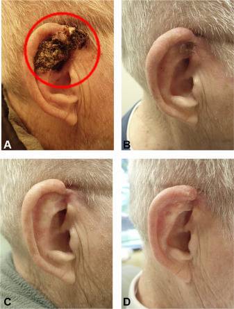
Left Image: Case Number Two (fig. 2 Yoon, (2021) showing large basal cell carcinoma right ear before treatment (Panel A, Red ellipse). Panel A, Pre-treatment. Panel B, 8 weeks of treatment. Panel C, 16 weeks of treatment. Panel D, 16 months after the initiation of therapy.
Conclusion: These two cases highlight the efforts of a local dermatologist to embrace integrative oncology concepts by combining two synergistic hedgehog inhibitor drugs to obtain greater efficacy with fewer side effects, with excellent results in these two cases. (1-42)
 Read more about using repurposed drugs to treat cancer in my book (left image) available on Amazon, Cracking Cancer Toolkit.
Jeffrey Dach MD
7450 Griffin Road, Suite 190
Davie, Fl 33314
954-792-4663
my blog: www.jeffreydachmd.com
References for Basal Cell Carcinoma
1) Yoon, Jaeyoung. “Vismodegib dose reduction effective when combined with itraconazole for the treatment of advanced basal cell carcinoma.” JAAD Case Reports 7 (2021): 107-109.
Limitations of vismodegib are the side effects experienced by most patients. Over half of the patients have mild-to-moderate adverse events, and 21.2% of the patients discontinued therapy due to this in the international, multicenter, single arm, phase II ERIVANCE BCC clinical trial by Genentech.4 The most common side effects are muscle spasm, fatigue, alopecia, dysgeusia, and weight loss.
An 84-year-old man was seen in the clinic for a BCC of the left chin (Fig 1, A), which recurred 2 years after Mohs micrographic surgery. He presented with a large, fixed, ulcerated, firm tumor. Magnetic resonance imaging showed the tumor approximating the bone. He refused surgery given his age and the possibility of an extensive surgical intervention. He was administered vismodegib 150 mg once per week and itraconazole 200 mg/day.
see photos
Notable clinical improvement was observed after 4 weeks of treatment (Fig 1, B). After 12 weeks, the area was completely healed, and the tissue was more supple (Fig 1, C). He continued the medications for a total of 28 weeks. His last follow-up was 16 months after the initiation of treatment, and there was no clinical evidence of tumor progression (Fig 1, D).
This patient experienced body hair loss and muscle spasms of the legs, which were described as mild and tolerable. There were no laboratory abnormalities throughout the treatment.
USE THIS Protocol for Pt w/basal cell metastatic
Case 2 – He was placed on
vismodegib 150 mg twice a week (Mondays and Fridays) and
itraconazole 100 mg/day.
The patient in case 2 denied any adverse effects at all.
An 85-year-old man presented for Mohs micrographic surgery for a large BCC on his right ear (Fig 2, A). He was concerned about the risk of deformity and asked for an alternative treatment. He was placed on vismodegib 150 mg twice a week (Mondays and Fridays) and itraconazole 100 mg/day.
Case 2 – He was placed on
vismodegib 150 mg twice a week (Mondays and Fridays) and itraconazole 100 mg/day. The patient in case 2 denied any adverse effects at all.
He was administered vismodegib 150 mg once per week and itraconazole 200 mg/day.
——————————–
2) Patil, Nitin Krishna, and Aditya Kumar Bubna. “Substantial reduction of basal cell carcinoma tumor size with itraconazole following treatment failure with intralesional 5-fluorouracil.” Journal of the Egyptian Women’s Dermatologic Society 20.1 (2023): 60-62.
The patient was then started on itraconazole (100 mg twice daily) for 8 months. At the end of 4 months, surface changes of the growth could be appreciated (Fig. 1d),
Kim et al. [4] in their report outlined clinical outcomes following institution of itraconazole monotherapy (dosing: 100 mg OD to 200 mg BD) for BCC for a period of 1–12 months. Of these, 57 primary BCCs in eight patients demonstrated a mean area reduction of 24%.
Another report from Poland elaborated slight clinical improvement following oral itraconazole monotherapy for locally advanced facial BCC in a 70-year-old man. Skin lesions in this patient stopped seeping, and
temporal ulceration had healed partially following 8 months of treatment with itraconazole [5].
Ip and McKerrow [6] though did not demonstrate profitability of itraconazole (200 mg/day) for cutaneous BCC. However, they did elucidate 30% reduction of pulmonary metastasis secondary to BCC, following itraconazole administration.
!!!!!!!!!!!!!!!!!!!!!!!!!!!!!!!!!!
YOON COmplete Regression
!!!!!!!!!!!!!!!!!!!!!!!!!!!!!!!!!!!
Yoon [7] reported complete regression of advanced facial BCC in two patients following low-dose vismodegib (150 mg once/twice per week) and itraconazole (100–200 mg/day).
After studying these reports, itraconazole definitely seems to hold promise as a new player for BCC therapy. However, one striking observation was that itraconazole monotherapy was ineffective in bringing about complete regression of the tumor, similar to our
finding. Only the report where vismodegib (low dose) and itraconazole were combined, was complete tumor remission obtained. So, whether itraconazole could be utilized as a valuable adjunct along with other treatments (medical and/or surgical) is something worth contemplating upon.
Currently, the exact duration of treatment with itraconazole for BCC has not been determined. Moreover, chronic administration of itraconazole, as well as likely adverse effects following its long-term intake, needs careful study. A report of Aspergillus spondylodiscitis in a patient with acute myeloid leukemia receiving 600–900 mg of itraconazole per day, however, elucidated a manageable toxicity profile [8].
At present, our patient is being maintained on itraconazole (100 mg Q12H), and even after a year of follow-up, there has neither been any increase or reduction in tumor size as when observed after 8 months of initiation of therapy. Besides, our patient is tolerating the drug well, without any untoward effects. Based on our findings, we suggest that itraconazole can be considered a second-line agent in certain cases of BCC, especially in those scenarios where access to surgery/radiotherapy (for BCC) is not available or when patients decline surgery or are unsuitable candidates for the same.
==========================================
3) Ip, Ken Hiu‐Kan, and Kevin McKerrow. “Itraconazole in the treatment of basal cell carcinoma: A case‐based review of the literature.” Australasian Journal of Dermatology 62.3 (2021): 394-397.
4) Li, Chun-Lan, et al. “Repurposed itraconazole for use in the treatment of malignancies as a promising therapeutic strategy.” Biomedicine & Pharmacotherapy 154 (2022): 113616.
5) Freitas, Raiza Dias, et al. “Inhibition of CAL27 oral squamous carcinoma cell by targeting hedgehog pathway with vismodegib or itraconazole.” Frontiers in Oncology 10 (2020): 563838.
6) El-Sheridy, Nabila A., et al. “Itraconazole for topical treatment of skin carcinogenesis: efficacy enhancement by lipid Nanocapsule formulations.” Journal of Biomedical Nanotechnology 18.1 (2022): 97-111.
7) Ramelyte, E., et al. “How to break resistance to hedgehog inhibitors in advanced basal cell carcinoma?.” British Journal of Dermatology 184.2 (2021): 359-361.
8) Svoboda, Steven A., Nathan M. Johnson, and Mariana A. Phillips. “Systemic Targeted Treatments for Basal Cell Carcinoma.” Cutis 109.6 (2022).
An open-label, exploratory phase 2 trial of 19 patients with BCC found that oral itraconazole 200 to 400 mg daily.
decreased tumor proliferative index by 45% (P=.04), as measured by Ki-67; SHH activity by 65% (P=.03), as measured by GLI1 messenger RNA; and mean tumor area by 24%.73 In a case series of 5 patients with mBCC refractory to conventional SHH inhibitor therapy, combined treatment with itraconazole and arsenic trioxide resulted in stable disease and a 75% reduction in SHH activity (P
Posaconazole is a second-generation antifungal agent that may serve as a potential alternative to itraconazole.77 Although clinical data are lacking, a basic science study
found that posaconazole could inhibit the growth of SHH-dependent BCC in vivo (in mice).78 Furthermore, posaconazole has demonstrated a better safety profile
with fewer and more mild side effects than itraconazole and does not require dose adjustment for those with hepatic or renal failure.79,80 Thus, posaconazole may be a
safer alternative to itraconazole for the treatment of BCC. Further clinical studies are needed to elucidate the potential synergistic effects of these antifungal agents with the
2 currently approved SHH inhibitors for the treatment of advanced BCC.
==============================
9) Marzagalli, Monica, et al. “Estrogen receptor β in melanoma: from molecular insights to potential clinical utility.” Frontiers in endocrinology 7 (2016): 140.
In vitro and in vivo antitumor effects on melanoma were also reported for 2-methoxyestradiol, an endogenous metabolite of estradiol; however, it must be pointed out that the antitumor activity of this compound was not found to be mediated by ERs (both α and β) activation (123, 124).
10) Dobos J, Timar J, Bocsi J, Burian Z, Nagy K, Barna G, et al. In vitro and in vivo
antitumor effect of 2-methoxyestradiol on human melanoma. Int J Cancer (2004) 112:771–6. doi:10.1002/ijc.20473
11) Ireson CR, Chander SK, Purohit A, Perera S, Newman SP, Parish D, et al.
Pharmacokinetics and efficacy of 2-methoxyoestradiol and 2-methoxyoestradiol-bis-sulphamate in vivo in rodents. Br J Cancer (2004) 90:932–7. doi:10.1038/sj.bjc.6601591
12) Chen, Baozhi, et al. “Posaconazole, a second-generation triazole antifungal drug, inhibits the hedgehog signaling pathway and progression of basal cell carcinoma.” Molecular cancer therapeutics 15.5 (2016): 866-876.
13) Bakshi, Anshika, et al. “Basal cell carcinoma pathogenesis and therapy involving hedgehog signaling and beyond.” Molecular carcinogenesis 56.12 (2017): 2543-2557.
14) Gambini, Donatella, et al. “Basal cell carcinoma and hedgehog pathway inhibitors: focus on immune response.” Frontiers in Medicine 9 (2022): 893063.
15) Cocuz, Iuliu Gabriel, et al. “Pathophysiology, Histopathology, and Differential Diagnostics of Basal Cell Carcinoma and Cutaneous Squamous Cell Carcinoma—An Update from the Pathologist’s Point of View.” International Journal of Molecular Sciences 25.4 (2024): 2220.
16) Bengoa-González, Alvaro, et al. “Advanced Periocular Basal Cell Carcinoma with Orbital Invasion: Update on Management and Treatment Advances.” Journal of Ophthalmology 2024.1 (2024): 4347707.
==============================
Fenofibrate
=================
O’Neill, W. Quinn, et al. “Repositioning fenofibrate to reactivate p53 and reprogram the tumor-immune microenvironment in HPV+ head and neck squamous cell carcinoma.” Cancers 14.2 (2022): 282.
Su, Tzu-Rong, et al. “Fenofibrate diminishes the self-renewal and metastasis potentials of oral carcinoma stem cells through NF-κB signaling.” Journal of the Formosan Medical Association 121.10 (2022): 1900-1907.
Fenofibrate OXPHOS inhibitor, 400 mg per day with evening meal.Plus Vitamin A, 25,000 to 50,000 iu/day
autophagy inhibitors enhance FF-induced glioblastoma cytotoxicity.
The combination of OXPHOS inhibitor fenofibrate with a GLYCOLYSIS inhibitor (such as DCA, diclofenac or quercetin), an autophagy inhibitor (such as hydroxychloroquine, loratidine, thymoquinone, etc.) and a microtubule inhibitor (such as mebendazole) might prove synergistic…
Be Careful with fenofibrate/Ivermectin combination both oxphas inhibitor with mitochonrial toxicity.
39) Jan, Chia-Ing, et al. “Fenofibrate suppresses oral tumorigenesis via reprogramming metabolic processes: potential drug repurposing for oral cancer.” International journal of biological sciences 12.7 (2016): 786.
40) Chang, Nai Wen, et al. “Fenofibrate exhibits a high potential to suppress the formation of squamous cell carcinoma in an oral-specific 4-nitroquinoline 1-oxide/arecoline mouse model.” Biochimica et Biophysica Acta (BBA)-Molecular Basis of Disease 1812.4 (2011): 558-564.
========================================
!!!!!!!!!!!!!!!!!!!!!!!!!!!!!! BEST !!!!!!!!!!!!!!!!!!!!!!!!!!!!!!!!!!!!!!!!!!
ER-Beta (Estriol-ER-Beta) (Premarin Bring Steroids ER-Beta) (Prometrium downregulates ER Alpha) (Androgens – 3BetaDiol)
41) Mancuso, M., et al. “Modulation of basal and squamous cell carcinoma by endogenous estrogen in mouse models of skin cancer.” Carcinogenesis 30.2 (2009): 340-347.
These and previous data from studies in rodents (30) suggest that estrogens may be key modulators of skin tumorigenesis. The effects of estrogens are mediated by estrogen receptor (ER)-a and ERb, members of the nuclear steroid receptor superfamily. Both ERs have been detected in the skin of rodents and humans, though with distinct expression patterns (36); specifically, ERb has been indicated as the predominant ER in human scalp skin (37), whereas in murine skin both ERs are expressed during hair follicle cycling in hair cycle-dependent manner (38).
In ovariectomized Ptch1þ/ and Car-S females, basal and squamous tumor induction were drastically increased over intact controls (CNs), and restored to levels observed in males, showing that endogenous estrogens play a critical role in protection against BCC and SCC carcinogenesis by diverse agents in mouse skin.
The skin locally synthesizes significant amounts of sexual hormones with intracrine or paracrine actions. However, the local level of each sexual steroid depends on the expression of androgen-and estrogen-synthesizing enzymes in different cell types (50). The role of estrogen in the regulation of hair follicle cycling in mice was rediscovered in the past decade, following a seminal paper by Oh et al. (33), showing that an ER pathway within the dermal papilla regulates the telogen–anagen follicle transition and that 17-b-estradiol blocks hair growth and arrests hair follicles in telogen.
Experimental data from the present study support the concept that
female sex hormones can be protective in non-melanoma skin carcinogenesis; in fact, we found that skin tumor development was significantly enhanced after ovarian hormone withdrawal in two independent experimental models. The results shown here demonstrate increased skin tumor incidence and multiplicity and decreased tumor latency in ovariectomized versus CN females, regardless of the nature of the keratinocyte-initiating agent (i.e. chemical for SCC and physical for BCC). Remarkably, malignant progression of benign papillomas to SCC occurred almost exclusively in OVX Car-S and was rare in CN females following two-stage carcinogenesis by DMBA/TPA.
To shed light on potential mechanisms involved in estrogen modulation of skin tumor progression, we examined ER protein levels in benign skin papillomas from the different Car-S groups. Immunoblots of papilloma extracts showed significantly increased expression of ERa and downregulation of ERb in tumors from OVX relative to
CN tumors, suggesting a role of the ratio ERa:ERb in susceptibility of skin to estrogen-modulated carcinogenesis, and a correlation of decreased ERb expression with increased malignant progression of initially benign papillomas in ovariectomized Car-S mice.
Previous studies have established a complex relationship between ERs and cyclin D1, with important implications for proliferation of estrogen-responsive tissues and deregulation of proliferation in cancer (42,43). To further explore this issue, we analyzed tumors for expression of cyclin D1, which among D-type cyclins controlling cell cycle regulation has been most directly implicated in oncogenesis. In the presence of estrogen, cyclin D1 is one important target gene through which estrogen-complexed ERa mediates its proliferative action, whereas estrogen-complexed ERb represses cyclin D1 gene transcription and blocks ERa-mediated induction when both receptors are present (56).
In the absence of estrogen, however, cyclin D1 is able to bind to and activate transcription mediated by ER-alpha (42,43,57). Significantly, we detected cyclin D1 upregulation in tumors from OVX relative to CN mice. Thus, our results suggest that in tumors from intact mice, where the ratio ERa:ERb is low, the protective role of ER-beta may be privileged over the proliferation stimulus mediated by the a-isoform, whereas in tumors from ovariectomized animals, the inverted ERa:ERb ratio may favor proliferation and malignant progression, possibly due to the oncogenic role of cyclin D1. This hypothesis is supported by the higher proliferation rate observed in papillomas from OVX compared with intact CN mice, a finding also observed in ER-positive breast cancer, where high cyclin D1 expression correlates with high Ki67 expression (58). We cannot exclude, however, that ovariectomy may modulate other factors involved in the regulation of skin development and functions, such as progesterone levels (59) and that this modulation may in turn influence tumor development.
In summary, our study shows for the first time a protective role of endogenous estrogen against basal and squamous skin tumorigenesis caused by physical or chemical agents in independent mouse models Finally, our study suggests that reciprocal expression of ERa and ERb may be associated with estrogen-mediated modulation of squamous epithelial carcinogenesis, with a key role played by cyclin D1.
=====================================================
—————————————
Cracking Cancer
Itraconazole, also known as Sporonox, is a common antifungal drug developed in the
1980s, usually prescribed as 100 mg or 200 mg oral capsules with daily dosage in the 100–600 mg range. Itraconazole is well tolerated when used long term to prevent or treat chronic fungal infection in immunosuppressed patients.
Four hundred milligrams per day for a year is not uncommon for chronic pulmonary aspergillosis or blastomycosis, both fungal infections.
Human dosage required to achieve anti-cancer levels used in animal studies is in the 600–900 mg per day range. (1–2)
Itraconazole has been in clinical use for 30 years with an established safety record.
Multiple phase 2 clinical trials investigating itraconazole for non-small-cell lung cancer,
prostate cancer, and basal cell carcinoma have been completed, showing an increase in progression- free and overall survival. (3–7)
Phase 2 Trial for Basal Skin Cancer Itraconazole
In 2014, a phase two trial of oral itraconazole for basal cell carcinoma was reported by
Dr. Daniel Kim et al., yielding impressive results in 19 patients placed into two groups receiving either 200 mg a day or 400 mg a day of the itraconazole drug for 1–3 months. Cancer cell proliferation was reduced by 65%, Hedgehog (Hh) activity reduced by 65%, and tumor area reduced by 24%. (56)
Both basal cell and squamous cell types of skin cancer show upregulation of the hedgehog/
GLI pathway. High expression of the Hh pathway is a prognostic factor that confers a
poor overall survival. The Hh/Gli pathway is inhibited by itraconazole, thus serving as an
effective anti-cancer agent for basal cell and squamous cell skin cancer. (56–59)
Jeffrey Dach MD
7450 Griffin Road, Suite 190
Davie, Fl 33314
954-792-4663
my blog: www.jeffreydachmd.com
Natural Thyroid Toolkit by Jeffrey Dach MD
Cracking Cancer Toolkit by Jeffrey Dach MD
Heart Book by Jeffrey Dach MD
www.naturalmedicine101.com
www.bioidenticalhormones101.com
www.truemedmd.com
www.drdach.com
Click Here for: Dr Dach’s Online Store for Pure Encapsulations Supplements
Click Here for: Dr Dach’s Online Store for Nature’s Sunshine Supplements
Web Site and Discussion Board Links:
jdach1.typepad.com/blog/
disc.yourwebapps.com/Indices/244066.html
disc.yourwebapps.com/Indices/244067.html
http://sci.med.narkive.com/covV2Qo2/jeffrey-dach-book-announcment-natural-medicine-101
The reader is advised to discuss the comments on these pages with his/her personal physicians and to only act upon the advice of his/her personal physician. Also note that concerning an answer which appears as an electronically posted question, I am NOT creating a physician — patient relationship. Although identities will remain confidential as much as possible, as I can not control the media, I can not take responsibility for any breaches of confidentiality that may occur.
Link to this Article
Copyright © 2024 Jeffrey Dach MD All Rights Reserved. This article may be reproduced on the internet without permission, provided there is a link to this page and proper credit is given. See Repost Guidelines.
FAIR USE NOTICE: This site contains copyrighted material the use of which has not always been specifically authorized by the copyright owner. We are making such material available in our efforts to advance understanding of issues of significance. We believe this constitutes a ‘fair use’ of any such copyrighted material as provided for in section 107 of the US Copyright Law. In accordance with Title 17 U.S.C. Section 107, the material on this site is distributed without profit to those who have expressed a prior interest in receiving the included information for research and educational purposes.
Serving Areas of: Hollywood, Aventura, Miami, Fort Lauderdale, Pembroke Pines, Miramar, Davie, Coral Springs, Cooper City, Sunshine Ranches, Hallandale, Surfside, Miami Beach, Sunny Isles, Normandy Isles, Coral Gables, Hialeah, Golden Beach ,Kendall,sunrise, coral springs, parkland,pompano, boca raton, palm beach, weston, dania beach, tamarac, oakland park, boynton beach, delray,lake worth,wellington,plantation
Published on October 30th, 2024 by Jeffrey Dach MD
The post Itraconazole and Vismodegib Combination for Basal Cell Carcinoma appeared first on Jeffrey Dach MD.
October 28, 2024
Estrogen Prevents Heart Disease Part Two
 Estrogen Prevents Heart Disease Part Two by Jeffrey Dach MD
In part one of this series, we discussed the history of hormone replacement regarding prevention of coronary artery disease finding a 50 percent reduction in mortality from heart disease depending on the study and the age of initiating estrogen replacement in post-menopausal women. The greatest cardiovascular benefit from HRT (hormone replacement therapy) was found in post-hysterectomy women with surgically induced menopause. Excellent cardiovascular mortality reductions of about 40 percent are obtained with HRT when started early, within 5 years of the menopausal transition. This is called the “Timing Hypothesis of Dr. Howard Hodis. Cardiovascular benefits of HRT are lost in women starting HRT more than 5 years after menopausal transition. (1)
This is part two. For part one, click here.
Header Image: Lab tech doing detection of bacterial lipopolysaccharides by electrophoresis. 2009 Author Aicomfoto CC 4.0 Courtesy of Wikimedia Commons.
How to Explain the Timing Hypothesis
The next logical question is how can we explain this “Timing Hypothesis?” What is the underlying pathophysiology that explains why estrogen replacement prevents coronary artery disease when started early, but not when started later?
Let us start by stating the obvious: the abrupt decline in estrogen levels in post-menopausal women triggers accelerated coronary artery disease, a degenerative change with calcified plaque formation in the wall of the coronary arteries. Once coronary plaque has been established, this degenerative change is difficult to reverse by starting estrogen later. This explanation doesn’t explain everything, and leaves important gaps in our understanding. Part two expands on the underlying pathophysiology of coronary artery disease in post-menopausal women and explains the “Timing Hypothesis” of Dr. Hodis.
Gut Permeability, Leaky Gut and Atherosclerosis
My 2018 book on coronary artery disease, entitled Heart Book, discusses the role of intestinal permeability, “Leaky Gut”, and low-level endotoxemia in the pathogenesis of coronary artery disease. The low-level endotoxemia arising from “leaky gut” leads to colonization of the arterial wall by polymicrobial biofilm which in turn leads to intense inflammatory reaction. The calcification within the arterial wall seen on the coronary calcium score is the body’s response to inflammation. Note: the coronary calcium score is a CAT scan test performed without IV contrast. The CT-angiogram test is a CAT scan performed with IV contrast.
In part one, we learned of two coronary calcium score studies derived from the WHI post menopausal patients showing the estrogen-treated group had lower calcium scores and reduced progression of calcium scores compared to the placebo-treated group. Coronary artery calcification is a response to polymicrobial infection in the wall of the artery, seeded from the gut microbiome and periodontal microorganisms. (2-3)
Heart Book, 2018
In my 2018 Heart Book, a mechanism was proposed to explain the train of events leading to coronary artery calcification. The originating event is “Leaky Gut”, a disruption of the mucosal barrier of the gut leading to leakage of LPS (lipopolysaccharide) and gram-negative microorganisms into the bloodstream, defined as low-level endotoxemia. Both LDL (low-density lipoprotein) and macrophages take up the LPS and whole micro-organisms, which then migrate into the arterial plaque, causing infection with polymicrobial biofilm. This incites an inflammatory reaction which leads to vascular calcification. Note: LPS is the outer coat of gram-negative bacteria, one of the most toxic substances known, and the cause of gram-negative septicemia, a dreaded complication with high mortality rate.(4)
Causes of Leaky Gut
There are many causes of “Leaky Gut.” The list includes fatty meals, high blood sugar (diabetes), dysbiosis with pathogenic bacteria, wheat gluten sensitivity which prolongs the opening of tight junctions, stress-altered permeability, etc. We can now add to this list the abrupt decline in estrogen levels which occurs at menopause. (5-6)
Menopausal Estrogen Decline Triggers Leaky Gut
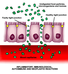 Left Image: Leaky Gut: The opening of intercellular tight junctions (increased intestinal permeability) allows uncontrolled passage of substances into the bloodstream, with consequent possible development of autoimmune and inflammatory diseases, infections, allergies or cancers, both intestinal and in other organs of the body.
April 2016 Author Ballena Blanca. Courtesy of
Wikimedia Commons.
The next obvious question is: can the sudden menopausal decline in estrogen trigger leaky gut and subsequent low-level endotoxemia? This is not well-known and not intuitively obvious. However, the medical literature on this is overwhelming, showing the menopausal decline in estrogen does indeed trigger disruption of the mucosal barrier of the gut, and causes increased gut permeability (i.e. Leaky Gut). We can now propose the menopausal decline in estrogen as the trigger for accelerated coronary atherosclerosis. Estrogen has two mechanisms for doing so. Firstly, estrogen is anti-inflammatory and secondly, estrogen controls and restores gut integrity. Anti-inflammatory effects of estrogen are mediated through the inhibition of Nuclear Factor Kappa B (NF-KB), the inflammatory master controller, which downregulates proinflammatory cytokines IL-1, IL-6, and TNF-alpha. Control of gut barrier integrity is mediated through ER-beta (estrogen receptor beta) which regulates tight junctions between epithelial cells of the gut lining. Thus, menopausal estrogen deficiency leads to disruption of the gut barrier, low-level endotoxemia, increased pro-inflammatory cytokines, and acceleration of atherosclerotic vascular disease, all reversed by estrogen replacement. In 2020, Dr. Albert Shieh studied animal models showing menopausal estrogen deficiency leads to increased gut permeability and inflammation, two major predicates for coronary artery disease and loss of bone mineral density (BMD), writing:
Inflammation is implicated in many aging-related disorders. In animal models, menopause [estrogen deficiency] leads to increased gut permeability and inflammation…Gut permeability increases during the MT [Menopausal Transition]. Greater gut permeability is associated with more inflammation and lower BMD [Bone Mineral Density]. Future studies should examine the longitudinal associations of gut permeability, inflammation, and BMD. Note: lower bone mineral density (BMD) is also associated with increased gut permeability and inflammation. (7-12)
In 2024, Dr. Xiuting Xiang studied the role of estrogen in the digestive system, noting ER-beta signaling regulates the permeability of the intestinal barrier, writing:
Overall, estrogen influences the composition and function of the gastrointestinal barrier and also impacts inflammatory processes within the digestive system. ER-beta signaling has been shown to regulate the permeability of the intestinal barrier by increasing the integrity of tight junctions through the expression of occludin and junctional adhesion molecule A. (13)
In 2024, Dr. Qinghai Meng from Nanjing, China found menopausal women have disordered gut microbiota which amplifies intestinal tight junction damage, accelerating atherosclerosis. Dr. Meng studied both women and a mouse model before and after menopause finding estrogen deficiency promotes microbiome disturbance, intestinal barrier damage, inflammation, and accelerates atherosclerosis, writing:
This study examined aortic estrogen receptor expression, histological changes, and gut microbiota in women before and after menopause, and tested serum estrogen levels, systemic inflammation, intestinal estrogen receptor expression, histological changes, atherosclerosis, and gut microbiota in low-density lipoprotein receptor knockout (LDLR-∕-) female mice before and after ovariectomy. We demonstrated that the downregulation of estrogen and estrogen receptors after menopause promotes gut microbiota disturbance in both women and female mice. We found that gut microbiota disturbance amplifies the intestinal barrier damage and aggravates systemic inflammation, thereby promoting atherosclerosis in female mice. Note: LDL receptor knockout mice are a good animal model for the study of atherosclerosis since they spontaneously develop atherosclerotic lesions. (14-16)
In 2023, Dr. Yuanuan Li used mice to show chronic psychological stress leads to decreased estrogen levels, disruption of the intestinal barrier with increased intestinal permeability, and increased pro-inflammatory cytokines. Estrogen replacement is protective and prevents these changes, writing:
Chronic psychological stress resulted in colonic mucosal injury, pro-inflammatory reaction, and decreased the diversity and richness of the colonic microbiota in pregnant mice. It was interesting that 25 pg/mL E2 provides better protective effect on intestinal epithelial cells…In summary, chronic stress significantly induces stress responses in maternal mice, leading to reduced estrogen secretion, disruption of the intestinal barrier, increased secretion of pro-inflammatory cytokines, decreased secretion of anti-inflammatory cytokines, and dysbiosis of the gut microbiota. Moreover, 25 pg/mL E2 protected the intestine. Our research demonstrates that maintaining stable maternal estrogen levels during pregnancy can safeguard intestinal health… (17)
Dr. Jane Yang on Estrogen and the GI Tract
In 2024, Dr. Jane Yang reviewed the entire medical literature on menopausal estrogen deficiency from 1972 to 2023. Within this larger topic, Dr. Jane Wang discusses estrogen’s importance for the gastrointestinal (GI) tract, pointing out the extensive presence of estrogen receptors throughout the GI tract. If you thought having estrogen receptors means estrogen is playing a controlling role, then you would be quite correct. ER-beta signalling controls the permeability of the gut barrier by maintaining the integrity of the tight junctions. When estrogen levels decline after menopause, this gut barrier integrity is lost, triggering leaky gut, low level endotoxemia, and accelerated atherosclerosis. Dr. Jane Wang writes:
ER-alpha, ER-beta, and GPER1 [G-protein coupled receptor 1] are found widely throughout the gastrointestinal (GI) tract…Overall, estrogen influences the composition and function of the gastrointestinal barrier and also impacts inflammatory processes within the digestive system. ER-beta signaling has been shown to regulate the permeability of the intestinal barrier by increasing the integrity of tight junctions through the expression of occludin and junctional adhesion molecule A… In multiple experimental models of colitis, estrogen has been shown to decrease intestinal inflammation and tissue damage. Note: GPER1 is the estrogen receptor on the cell membrane involved in immediate signaling. The other two receptors must enter the nucleus which takes more time. (18-26)
Menopause as an Inflammatory Event
There is considerable evidence for viewing the menopausal transition as an inflammatory event. In 2007, Dr. Toshiyuki Yasui found that serum IL-6, the main inflammatory cytokine was inversely correlated with serum estradiol levels. As estradiol decreased, the pro-inflammatory cytokines increased. In 2021, Dr. Haidong Wang found estradiol has an inhibitory effect on NF-KappaB, the inflammatory master controller. (27-29)
In 2005, Dr Serena Ghisletti studied the mechanism by which estradiol exerts its anti-inflammatory effects, finding estrogen blocks the p65 transcription factor (a member of the NF-KappaB family), inhibiting the p65 intracellular transport to the nucleus. This anti-inflammatory effect of estrogen is mediated through ER-alpha, writing:
Estrogen is an immunoregulatory agent, in that hormone deprivation increases while 17β-estradiol (E2) administration blocks the inflammatory response; however, the underlying mechanism is still unknown. The transcription factor p65/relA, a member of the nuclear factor κB (NF-κB) family, plays a major role in inflammation and drives the expression of proinflammatory mediators. Here we report a novel mechanism of action of E2 in inflammation. We observe that in macrophages E2 blocks lipopolysaccharide-induced DNA binding and transcriptional activity of p65 by preventing its nuclear translocation. This effect is selectively activated in macrophages to prevent p65 activation by inflammatory agents and extends to other members of the NF-κB family, including c-Rel and p50. We observe that E2 activates a rapid and persistent response that involves the activation of phosphatidylinositol 3-kinase, without requiring de novo protein synthesis or modifying Iκ-Bα degradation and mitogen-activated protein kinase activation. Using a time course experiment and the microtubule-disrupting agent nocodazole, we observe that the hormone inhibits p65 intracellular transport to the nucleus. This activity is selectively mediated by estrogen receptor alpha (ER-alpha) and not ERβ and is not shared by conventional anti-inflammatory drugs. These results unravel a novel and unique mechanism for E2 anti-inflammatory activity, which may be useful for identifying more selective ligands for the prevention of the inflammatory response. (30-35)
In 2020, Dr. Micheline McCarthy reviewed the menopausal transition as an inflammatory event, suggesting the menopausal decline in estrogen levels drives a systemic pro-inflammatory state, writing:
It is now known that one of the key functions of estrogen is to work as a potent anti-inflammatory factor …The presence of the inflammasome complex in the cerebrospinal fluid of post-menopausal women suggests that the decline in estrogens induces a pro-inflammatory state...There is increasing and compelling evidence showing that estrogen decline during the menopausal transition drives a systemic inflammatory state. This state is characterized by systemic pro-inflammatory cytokines derived from reproductive tissues, alteration in the cellular immune profile, increased availability of inflammasome proteins in the CNS, and a pro-inflammatory microenvironment which makes the brain more susceptible to ischemic and other stressors.… The use of ER-beta-selective agonists may constitute a safer and more effective target for future therapeutic research than an ER-alpha agonist or E2 [estradiol]. ER-beta activation in the brain confers ischemic protection, stimulates mitochondrial functions, and inhibits inflammasome activation. ER-beta agonists may be safer in that ER-beta lacks the ability to stimulate the proliferation of breast or endometrial tissue. The ER-beta agonist may be able to act both on the cerebro- and cardiovascular system to reduce the ischemic burden. Thus, ER-beta signaling is a guide for future translational research to reduce cognitive decline and cerebral ischemia incidents and impact in post-menopausal women, while avoiding the side effects produced by chronic E2 [estradiol] treatment…Emerging evidence is showing that peri-menopause is pro-inflammatory and disrupts estrogen-regulated neurological systems. Estrogen is a master regulator that functions through a network of estrogen receptors subtypes alpha (ER-α) and beta (ER-β). Estrogen receptor-beta has been shown to regulate a key component of the innate immune response known as the inflammasome, and it also is involved in regulation of neuronal mitochondrial function. (36-38)
Low Grade Endotoxemia, LPS and Infection in Atherosclerotic Plaque
In 2023, Dr. Francesco Violi reviewed the link between gut-derived low grade endotoxemia, atherosclerosis and cardiovascular disease. Gut dysbiosis (Leaky Gut) is implicated in atherosclerosis via leakage of live bacteria and LPS into the blood stream causing low-level endotoxemia. Studies show the presence of LPS adjacent to macrophages within atherosclerotic plaques. Dr. Francesco Violi writes:
A growing body of evidence indicates that gut dysbiosis is implicated in the atherothrombotic process via increased translocation of viable bacteria or bacterial products such as lipopolysaccharides (LPS) and trimethylamine-N-oxide (TMAO) into the systemic circulation ….Support for the putative role of LPS in atherosclerosis has been provided by immunohistochemistry analysis of carotid atherosclerotic plaques from patients undergoing endarterectomy, which revealed the presence of LPS adjacent to plaque macrophages with high TLR4 levels. By contrast, LPS was not detected in atherosclerosis-free thyroid arteries from the same patients….Experimental data support the association between low-grade endotoxaemia, atherosclerosis and thrombosis, and indicate that gut dysbiosis-induced changes in intestinal permeability are a key step for LPS translocation into the systemic circulation. (39)
In 2011, Dr. Omry Koren 16s ribosomal RNA sequencing to find bacterial DNA present within atherosclerotic plaques, writing:
Using qPCR, we show that bacterial DNA was present in the atherosclerotic plaque and that the amount of DNA correlated with the amount of leukocytes in the atherosclerotic plaque. To investigate the microbial composition of atherosclerotic plaques and test the hypothesis that the oral or gut microbiota may contribute to atherosclerosis in humans, we used 454 pyrosequencing of 16S rRNA genes to survey the bacterial diversity of atherosclerotic plaque, oral, and gut samples of 15 patients with atherosclerosis, and oral and gut samples of healthy controls…Our analysis also revealed several OTUs [operational taxonomic units] shared between the atherosclerotic plaque and the gut, suggesting that bacteria present in the atherosclerotic plaque could also be derived from the distal gut as well as the oral cavity. One mechanism by which bacteria could reach the atherosclerotic plaque is phagocytosis by macrophages at epithelial linings (e.g., the oral cavity, gut, and the lung). Upon phagocytosis, the macrophages become activated, and when they reach the activated endothelium of the atheroma, they leave the blood stream to enter the atheroma and transform into cholesterol-laden foam cells. In support of this mechanism, patients with cardiovascular disease have a twofold increase of C. pneumonia-infected peripheral blood mononuclear cells compared with controls. Furthermore, bacteria are only present in atheromas and not in healthy aortic tissues in mice and have been identified in human atherosclerotic plaques. Thus, infected macrophages may specifically target bacteria to atheromas… In summary, we detected key bacterial members of dental plaque in atherosclerotic plaques in humans, as well as a novel common member, Chryseomonas, in all atherosclerotic plaques. In addition, the atherosclerotic plaques contained numerous bacteria from different phyla. Our findings strongly support the hypothesis that the oral cavity and gut can be sources for atherosclerotic plaque-associated bacteria. (40)
In 2018, Dr. Roberto Canevale found LPS from E. Coli (gram negative bacteria) within atherosclerotic plaque material. (41-42)
In 2022, Dr. Iman Razeghian-Jahromi reviewed the medical literature on the prevalence of micro-organisms within atherosclerotic plaques of the coronary arteries finding 44 studies, writing:
In this systematic review and meta-analysis, the existence of pathogens in atherosclerotic plaques of coronary arteries was investigated in CAD patients…44 studies were selected…Infection of vascular cells, detection of the microbes within the atherosclerotic plaque, and the development of atherosclerotic lesions after microbial infection in animal models reinforce the direct association of infection with atherosclerosis….Our studies show the presence of different pathogens (bacteria and virus) in the atherosclerotic plaques of coronary arteries...This may show the implication of a microbial center for the initiation and development of atherosclerotic plaque…Several studies reported the presence of more than one pathogen in atherosclerotic plaques. The simultaneous existence of some bacteria synergistically enhances their virulence. Virulence factors of bacteria including fimbriae, degradative enzymes, exopolysaccharide capsules, toxins, and atypical lipopolysaccharides trigger the process of inflammation and affect vital organs like the cardiovascular system. These adverse effects will be more detrimental in mixed infections…Atherosclerosis may be originated from bacterial infection in terms of microbial symbiosis and inflammatory stimulus]. Microbial agents contribute to the atherosclerosis process directly by infecting the vascular cells or indirectly by activation of inflammatory cytokines. … It seems that the entry of bacteria and other microbiome populations into the bloodstream is a continuous flow which inevitably leads to a surge in the expression of inflammatory cytokines and chemokines. These factors could be drivers of CAD [71]. For example, it was shown that bacterial lipopolysaccharide (LPS) upregulates LDL levels, increasing the risk of CAD. Indeed, the remnants of bacteria like DNA or membrane phospholipids provoke CAD via modulating adipose or vascular tissues. However, it was reported that the development of atherosclerotic lesions is largely accelerated by live organisms rather than heat-killed ones or their LPS. Reports on successful culturing of pathogens after their isolation from atheroma only existed about C. pneumoniae and E. hormaechei. (43-57)
Conclusion: When one begins to understand the role of leaky gut, low level endotoxemia and microbial colonization of atherosclerotic plaque, one begins to realize the role of estrogen in prevention of coronary artery disease. Firstly. estrogen is an anti-inflammatory agent, and secondly, estrogen maintains the gut barrier. Menopausal loss of estrogen increases systemic inflammation, disruption of the gut barrier, low level endotoxemia and atherosclerosis. It becomes clear these two roles of estrogen are in fact what is preventing the acute onset of coronary artery disease in the post menopausal time frame. It also becomes clear why lowering serum cholesterol with statin drugs fails in primary prevention. Although statin drugs are anti-inflammatory, and antimicrobial, two pleiotropic effects, statin drugs do not address the gut membrane barrier, while menopausal estrogen replacement does. Another difference is the well-known adverse side effects of statins which are not shared by hormone replacement, namely, muscle pain, cognitive dysfunction, and neuropathy.
The above proposed mechanism of leaky gut and low level endotoxemia also explains the timing hypothesis of Dr. Howard Hodis. Promptly starting estrogen replacement at the onset of menopausal symptoms prevents the detrimental effects on the gut barrier, prevents low level endotoxemia, and microbial infiltration of the atherosclerotic plaque. This is true prevention for coronary artery disease. The most casual observer can see the overwhelming evidence for the infection theory of coronary artery disease, yet the cholesterol theory is too entrenched and the financial stakes are too high for mainstream medicine to make the paradigm shift. Mainstream medicine clings to the old dogmas, and ignores and refuses to accept all the studies listed here that prove microbial infection is the over-riding pathology in the coronary artery plaque which causes calcification seen on the Calcium Score, a CAT scan test. Instead of prescribing hormone replacement, the menopausal woman is handed out statin drugs nonchalantly by the primary care and cardiologist, oblivious to the fact statin drugs are ineffective for primary prevention of coronary artery disease, and are associated with the horrendous adverse side effects of muscle pain, cognitive dysfunction, and neuropathy. For more on this see the Heart Book (2018) by Jeffrey Dach MD. (4)
 My book is called: Heart Book
If you liked this article, you may like my book, entitled Heart Book on Amazon. See cover image at left.
Click Here for Link to book on Amazon
Articles with Related Interest:
Estrogen Prevents Heart Disease Part One
All Bioidentical Hormone Articles
Jeffrey Dach MD
7450 Griffin Road, Suite 190
Davie, Fl 33314
954-792-4663
my blog: www.jeffreydachmd.com
Natural Thyroid Toolkit by Jeffrey Dach MD
Cracking Cancer Toolkit by Jeffrey Dach MD
Heart Book by Jeffrey Dach MD
References for Chapter 10 Estrogen Prevents Heart Disease Part Two
1) Hodis, Howard N., and Wendy J. Mack. “Menopausal hormone replacement therapy and reduction of all-cause mortality and cardiovascular disease: it is about time and timing.” The Cancer Journal 28.3 (2022): 208-223.
2) Manson, JoAnn E., et al. “Estrogen therapy and coronary-artery calcification.” New England Journal of Medicine 356.25 (2007): 2591-2602.
3) Budoff, Matthew J., et al. “Effects of hormone replacement on progression of coronary calcium as measured by electron beam tomography.” Journal of women’s health 14.5 (2005): 410-417.
4) Dach, Jeffrey, “Heart Book”, Medical Muse Press, August 2, 2018.
5) Camilleri, Michael. “Leaky gut: mechanisms, measurement and clinical implications in humans.” Gut 68.8 (2019): 1516-1526.
6) Stewart, Amy Stieler, Shannon Pratt-Phillips, and Liara M. Gonzalez. “Alterations in intestinal permeability: the role of the “leaky gut” in health and disease.” Journal of equine veterinary science 52 (2017): 10-22.
7) Shieh, Albert, et al. “Gut permeability, inflammation, and bone density across the menopause transition.” JCI insight 5.2 (2020).
8) Ricardo-da-Silva, Fernanda Yamamoto, et al. “Estradiol prevented intestinal ischemia and reperfusion-induced changes in intestinal permeability and motility in male rats.” Clinics 76 (2021): e2683.
9) Li, Yuanyuan, et al. “Chronic stress that changed intestinal permeability and induced inflammation was restored by estrogen.” International Journal of Molecular Sciences 24.16 (2023): 12822.
10) Acharya, Kalpana D., et al. “Distinct changes in gut microbiota are associated with estradiol-mediated protection from diet-induced obesity in female mice.” Metabolites 11.8 (2021): 499.
11) Fidya, et al. “Protective role of estrogen through G-protein coupled receptor 30 in a colitis mouse model.” Histochemistry and Cell Biology 161.1 (2024): 81-93.
12) Jin, Ye, et al. “Estrogen deficiency aggravates fluoride-induced small intestinal mucosa damage and junctional complexes proteins expression disorder in rats.” Ecotoxicology and Environmental Safety 246 (2022): 114181.
13) Xiang, Xiuting, Praneetha Palasuberniam, and Rahmawati Pare. “The Role of Estrogen across Multiple Disease Mechanisms.” Current Issues in Molecular Biology 46.8 (2024): 8170-8196.
14) Meng, Qinghai, et al. “Disordered gut microbiota in postmenopausal stage amplifies intestinal tight junction damage to accelerate atherosclerosis.” Beneficial Microbes 1.aop (2024): 1-23.
15) Meng, Qinghai, et al. “The gut microbiota during the progression of atherosclerosis in the perimenopausal period shows specific compositional changes and significant correlations with circulating lipid metabolites.” Gut Microbes 13.1 (2021): 1880220.
16) Brandsma, Eelke, et al. “A proinflammatory gut microbiota increases systemic inflammation and accelerates atherosclerosis.” Circulation research 124.1 (2019): 94-100.
17) Li, Yuanyuan, et al. “Chronic stress that changed intestinal permeability and induced inflammation was restored by estrogen.” International Journal of Molecular Sciences 24.16 (2023): 12822.
18) Yang, Jane L., et al. “Estrogen deficiency in the menopause and the role of hormone therapy: integrating the findings of basic science research with clinical trials.” Menopause (2024): 10-1097.
19) Braniste, Viorica, et al. “Oestradiol decreases colonic permeability through estrogen receptor beta‐mediated up‐regulation of occludin and junctional adhesion molecule‐A in epithelial cells.” The Journal of physiology 587.13 (2009): 3317-3328.
20) Song, Chin-Hee, et al. “Effects of 17β-estradiol on colonic permeability and inflammation in an azoxymethane/dextran sulfate sodium-induced colitis mouse model.” Gut and liver 12.6 (2018): 682.
21) Looijer-van Langen, Mirjam, et al. “Estrogen receptor-beta signaling modulates epithelial barrier function.” American Journal of Physiology-Gastrointestinal and Liver Physiology 300.4 (2011): G621-G626.
22) Collins, Fraser L., et al. “Temporal and regional intestinal changes in permeability, tight junction, and cytokine gene expression following ovariectomy‐induced estrogen deficiency.” Physiological reports 5.9 (2017): e13263.
23) Zhou, Zejun, et al. “Progesterone decreases gut permeability through upregulating occludin expression in primary human gut tissues and Caco-2 cells.” Scientific reports 9.1 (2019): 8367.
24) Pigrau, M., et al. “The joint power of sex and stress to modulate brain–gut–microbiota axis and intestinal barrier homeostasis: implications for irritable bowel syndrome.” Neurogastroenterology & Motility 28.4 (2016): 463-486.
25) van der Giessen, Janine, et al. “A direct effect of sex hormones on epithelial barrier function in inflammatory bowel disease models.” Cells 8.3 (2019): 261.
26) Roomruangwong, Chutima, et al. “The menstrual cycle may not be limited to the endometrium but also may impact gut permeability.” Acta Neuropsychiatrica 31.6 (2019): 294-304.
27) Yasui, Toshiyuki, et al. “Changes in serum cytokine concentrations during the menopausal transition.” Maturitas 56.4 (2007): 396-403.
28) Wang, Haidong, et al. “17β-Estradiol alleviates intervertebral disc degeneration by inhibiting NF-κB signal pathway.” Life Sciences 284 (2021): 119874.
29) Wen, Yi, et al. “Estrogen attenuates nuclear factor-kappa B activation induced by transient cerebral ischemia.” Brain research 1008.2 (2004): 147-154.
30) Ghisletti, Serena, et al. “17β-estradiol inhibits inflammatory gene expression by controlling NF-κB intracellular localization.” Molecular and cellular biology 25.8 (2005): 2957-2968.
31) Xing, Dongqi, et al. “Estrogen modulates NFκB signaling by enhancing IκBα levels and blocking p65 binding at the promoters of inflammatory genes via estrogen receptor-β.” PloS one 7.6 (2012): e36890.
32) Liu, Hui, Kenian Liu, and Donald L. Bodenner. “Estrogen receptor inhibits interleukin-6 gene expression by disruption of nuclear factor κB transactivation.” Cytokine 31.4 (2005): 251-257.
33) Okamoto, Mariko, et al. “The membrane‐type estrogen receptor G‐protein‐coupled estrogen receptor suppresses lipopolysaccharide‐induced interleukin 6 via inhibition of nuclear factor‐kappa B pathway in murine macrophage cells.” Animal Science Journal 88.11 (2017): 1870-1879.
33) Deshpande, Rohini, et al. “Estradiol down‐regulates LPS‐induced cytokine production and NFkB activation in murine macrophages.” American journal of reproductive immunology 38.1 (1997): 46-54.
34) Zang, Ying CQ, et al. “Regulatory effects of estriol on T cell migration and cytokine profile: inhibition of transcription factor NF-κB.” Journal of neuroimmunology 124.1-2 (2002): 106-114.
35) Vegeto, Elisabetta, et al. “Estrogen receptor-α mediates the brain antiinflammatory activity of estradiol.” Proceedings of the National Academy of Sciences 100.16 (2003): 9614-9619.
36) McCarthy, Micheline, and Ami P. Raval. “The peri-menopause in a woman’s life: a systemic inflammatory phase that enables later neurodegenerative disease.” Journal of neuroinflammation 17 (2020): 1-14.
37) Zhang, Wen-yuan, et al. “Neuroprotective effects of vitamin D and 17ß-estradiol against ovariectomy-induced neuroinflammation and depressive-like state: Role of the AMPK/NF-κB pathway.” International Immunopharmacology 86 (2020): 106734.
38) Son, Hee Jin, et al. “17β-Estradiol reduces inflammation and modulates antioxidant enzymes in colonic epithelial cells.” The Korean Journal of Internal Medicine 35.2 (2020): 310.
39) Violi, Francesco, et al. “Gut-derived low-grade endotoxaemia, atherothrombosis and cardiovascular disease.” Nature reviews cardiology 20.1 (2023): 24-37.
40) Koren, Omry, et al. “Human oral, gut, and plaque microbiota in patients with atherosclerosis.” Proceedings of the National Academy of Science. Vol. 108. 2011.
41) Carnevale, Roberto, et al. “Localization of lipopolysaccharide from Escherichia Coli into human atherosclerotic plaque.” Scientific Reports 8 (2018).
42) Wiedermann CJ, et al. Association of endotoxemia with carotid atherosclerosis and cardiovascular disease: prospective results from the Bruneck study. J. Am. Coll. Cardiol. 1999;34:1975–1981.
43) Razeghian-Jahromi, Iman, et al. “Prevalence of Microorganisms in Atherosclerotic Plaques of Coronary Arteries: A Systematic Review and Meta‐Analysis.” Evidence‐Based Complementary and Alternative Medicine 2022.1 (2022): 8678967.
44) Khan, Ikram, et al. “Analysis of the blood bacterial composition of patients with acute coronary syndrome and chronic coronary syndrome.” (2022).
45) Cretoiu, Dragos, et al. “Gut microbiome, functional food, atherosclerosis, and vascular calcifications—Is there a missing link?.” Microorganisms 9.9 (2021): 1913.
46) Pisano, Eugenia, et al. “Microbial signature of plaque and gut in acute coronary syndrome.” Scientific Reports 13 (2023).
47) Joshi, Chaitanya, et al. “Detection of periodontal microorganisms in coronary atheromatous plaque specimens of myocardial infarction patients: A systematic review and meta-analysis.” Trends in cardiovascular medicine 31.1 (2021): 69-82.
48) Rao, Amita, et al. “Molecular analysis shows the presence of periodontal bacterial DNA in atherosclerotic plaques from patients with coronary artery disease.” Indian Heart Journal 73.2 (2021): 218-220.
49) Jonsson, Annika Lindskog, et al. “Bacterial profile in human atherosclerotic plaques.” Atherosclerosis 263 (2017): 177-183.
50) Pavlic, Verica, et al. “Identification of periopathogens in atheromatous plaques obtained from carotid and coronary arteries.” BioMed Research International 2021.1 (2021): 9986375.
51) Masoumi, Omid, et al. “Detection of fungal elements in atherosclerotic plaques using mycological, pathological and molecular methods.” Iranian Journal of Public Health 44.8 (2015): 1121.
52) Sato, Ayako, et al. “Metagenomic Analysis of Bacterial Microflora in Dental and Atherosclerotic Plaques of Patients With Internal Carotid Artery Stenosis.” Clinical Medicine Insights: Cardiology 18 (2024): 11795468231225852.
53) Ravnskov, Uffe, Abdullah Alabdulgader, and Kilmer S. McCully. “Infections may cause arterial inflammation, atherosclerosis, myocarditis and cardiovascular disease.” Medical Research Archives 11.5 (2023).
54) Lv, Hang, et al. “Characteristics of the gut microbiota of patients with symptomatic carotid atherosclerotic plaques positive for bacterial genetic material.” Frontiers in Cellular and Infection Microbiology 13 (2023).
55) Kissinger, Dohn. “Are bacterial infections a major cause of cardiovascular disease?.” Frontiers in Cardiovascular Medicine 11 (2024): 1389109.
56) Huang, Xiaofei, et al. “The Roles of Periodontal Bacteria in Atherosclerosis.” International Journal of Molecular Sciences 24.16 (2023): 12861.
57) Jayasinghe, Thilini N., et al. “Identification of oral bacteria in the gut, atherosclerotic plaque, and cultured blood samples of patients with cardiovascular diseases–A secondary analysis of metagenomic microbiome data.” (2023).
=======================================================
Jeffrey Dach MD
7450 Griffin Road, Suite 190
Davie, Fl 33314
954-792-4663
my blog: www.jeffreydachmd.com
Natural Thyroid Toolkit by Jeffrey Dach MD
Cracking Cancer Toolkit by Jeffrey Dach MD
Heart Book by Jeffrey Dach MD
www.naturalmedicine101.com
www.bioidenticalhormones101.com
www.truemedmd.com
www.drdach.com
Click Here for: Dr Dach’s Online Store for Pure Encapsulations Supplements
Click Here for: Dr Dach’s Online Store for Nature’s Sunshine Supplements
Web Site and Discussion Board Links:
jdach1.typepad.com/blog/
disc.yourwebapps.com/Indices/244066.html
disc.yourwebapps.com/Indices/244067.html
http://sci.med.narkive.com/covV2Qo2/jeffrey-dach-book-announcment-natural-medicine-101
The reader is advised to discuss the comments on these pages with his/her personal physicians and to only act upon the advice of his/her personal physician. Also note that concerning an answer which appears as an electronically posted question, I am NOT creating a physician — patient relationship. Although identities will remain confidential as much as possible, as I can not control the media, I can not take responsibility for any breaches of confidentiality that may occur.
Link to this Article
Copyright © 2024 Jeffrey Dach MD All Rights Reserved. This article may be reproduced on the internet without permission, provided there is a link to this page and proper credit is given. See Repost Guidelines.
FAIR USE NOTICE: This site contains copyrighted material the use of which has not always been specifically authorized by the copyright owner. We are making such material available in our efforts to advance understanding of issues of significance. We believe this constitutes a ‘fair use’ of any such copyrighted material as provided for in section 107 of the US Copyright Law. In accordance with Title 17 U.S.C. Section 107, the material on this site is distributed without profit to those who have expressed a prior interest in receiving the included information for research and educational purposes.
Serving Areas of: Hollywood, Aventura, Miami, Fort Lauderdale, Pembroke Pines, Miramar, Davie, Coral Springs, Cooper City, Sunshine Ranches, Hallandale, Surfside, Miami Beach, Sunny Isles, Normandy Isles, Coral Gables, Hialeah, Golden Beach ,Kendall,sunrise, coral springs, parkland,pompano, boca raton, palm beach, weston, dania beach, tamarac, oakland park, boynton beach, delray,lake worth,wellington,plantation
Published on October 28th, 2024 by Jeffrey Dach MD
The post Estrogen Prevents Heart Disease Part Two appeared first on Jeffrey Dach MD.
October 11, 2024
Menopausal Hormone Therapy Podcast with Dr. Amy Brenner
 Menopausal Hormone Replacement Therapy with Drs. Amy Brenner and Jeffrey Dach
Thanks to Dr. Amy Brenner for having me on her podcast for a discussion of menopausal hormone replacement.
Link to Dr. Amy Brenner Web Site.
Amy Brenner MD and Associates
6413 Thornberry Ct.
Mason, OH 45040
tel:+15137700787
Scroll Down to Watch the Part One and Part Two of the Podcast:
=============================================================
=================================
Dr. Amy Brenner Podcast Part Two
Menopausal HRT: Fact v Fantasy with Dr. Jeffrey Dach Pt 2 HealthiHer Podcast #94
In the first part of “Menopausal Hormone Replacement Therapy: Fact v. Fantasy” our hormone specialists Dr. Amy Brenner and Dr. Jeffrey Dach tackle the myths and truths surrounding menopausal hormone therapy. If you’ve ever wondered about the safety and benefits of hormone replacement during menopause, this episode is for you.
Dr. Brenner and Dr. Dach address the core concerns and misconceptions about hormone therapy during menopause. Why do some women fear hormone replacement therapy (HRT)? Are these fears rooted in fact or fiction? They discuss whether estrogen, progesterone, and testosterone impact breast cancer risk, helping to clarify whether these hormones contribute to or protect against cancer. They thoroughly evaluate studies and present the finds during the episode to help bring evidence into perspective.
Dr. Dach and Dr. Brenner discuss the confusion between synthetic progestins vs. progesterone and the risk of developing breast cancer and what you should consider if you start taking progesterone.
Additionally, Dr. Dach touches on certain genetic conditions that may impact some women’s probability of developing breast cancer.
Join us for a comprehensive exploration of menopausal hormone therapy, where Dr. Brenner and Dr. Dach separate fact from fiction, providing you with the essential information to make well-informed decisions about your health during menopause.
In Part Two (below) , the next episode , the experts will discuss certain practices you can introduce to protect your breast health and the experts also delve into how hormone therapy during menopause influences overall health beyond cancer risks. Does menopausal hormone therapy reduce mortality, prevent osteoporosis, heart disease, and dementia? Learn how these therapies can affect your long-term health and quality of life.
In the second part of Menopausal Hormone Replacement Therapy: Fact V. Fantasy, Dr. Jeffrey Dach and Dr. Amy Brenner continue their discussion on HRT and the misrepresentations surrounding these treatments. Join the two hormone experts as they discuss how to protect your breast health as you age and diving further into the discussion regarding progesterone usage versus progestins.
========================================
Jeffrey Dach MD
7450 Griffin Road, Suite 190
Davie, Fl 33314
954-792-4663
my blog: www.jeffreydachmd.com
Natural Thyroid Toolkit by Jeffrey Dach MD
Cracking Cancer Toolkit by Jeffrey Dach MD
Heart Book by Jeffrey Dach MD
www.naturalmedicine101.com
www.bioidenticalhormones101.com
www.truemedmd.com
www.drdach.com
Click Here for: Dr Dach’s Online Store for Pure Encapsulations Supplements
Click Here for: Dr Dach’s Online Store for Nature’s Sunshine Supplements
Web Site and Discussion Board Links:
jdach1.typepad.com/blog/
disc.yourwebapps.com/Indices/244066.html
disc.yourwebapps.com/Indices/244067.html
http://sci.med.narkive.com/covV2Qo2/jeffrey-dach-book-announcment-natural-medicine-101
The reader is advised to discuss the comments on these pages with his/her personal physicians and to only act upon the advice of his/her personal physician. Also note that concerning an answer which appears as an electronically posted question, I am NOT creating a physician — patient relationship. Although identities will remain confidential as much as possible, as I can not control the media, I can not take responsibility for any breaches of confidentiality that may occur.
Link to this Article
Copyright © 2024 Jeffrey Dach MD All Rights Reserved. This article may be reproduced on the internet without permission, provided there is a link to this page and proper credit is given. See Repost Guidelines.
FAIR USE NOTICE: This site contains copyrighted material the use of which has not always been specifically authorized by the copyright owner. We are making such material available in our efforts to advance understanding of issues of significance. We believe this constitutes a ‘fair use’ of any such copyrighted material as provided for in section 107 of the US Copyright Law. In accordance with Title 17 U.S.C. Section 107, the material on this site is distributed without profit to those who have expressed a prior interest in receiving the included information for research and educational purposes.
Serving Areas of: Hollywood, Aventura, Miami, Fort Lauderdale, Pembroke Pines, Miramar, Davie, Coral Springs, Cooper City, Sunshine Ranches, Hallandale, Surfside, Miami Beach, Sunny Isles, Normandy Isles, Coral Gables, Hialeah, Golden Beach ,Kendall,sunrise, coral springs, parkland,pompano, boca raton, palm beach, weston, dania beach, tamarac, oakland park, boynton beach, delray,lake worth,wellington,plantation
Published on October 11th, 2024 by Jeffrey Dach MD
The post Menopausal Hormone Therapy Podcast with Dr. Amy Brenner appeared first on Jeffrey Dach MD.
September 13, 2024
Florida Department of Health Advises Against C0\/lD B00STERS

Florida Dept of Health, Surgeon General, Joseph Ladapo, MD, Advises Against Use of mRNA C0\/lD \/a<
Updated Guidance for C0\/lD \/a<
Tallahassee, Fla. – The Florida Department of Health (Department) is reminding health care providers of the importance of remaining up to date with current literature related to C0\/lD \/a< and b00sters, and the importance of providing patients with informed consent. Click Here for Original Version.
On August 22, 2024, the United States Food and Drug Administration (FDA) approved and authorized updated versions of mRNA C0\/lD \/a< from Pfizer-BioNtech and Moderna. The FDA approved the C0\/lD \/a< for people 12 and older and provided emergency use authorization for children 6 months to 11 years old. The stated target of these boosters is the Omicron variant which is not causing a significant number of infections.
No Clinical Trial Data, No Protection for Current Dominant Strain
The most recent booster approval was granted in the absence of booster-specific clinical trial data performed in humans. Furthermore, this booster does not protect against the currently dominant strain, accounting for approximately 37% of infections in the United States. There are currently limited data to inform whether these boosters offer any substantial protection against the virus and subsequent circulating variants. Although randomized clinical trials are normally used to approve therapeutics, the federal government has not required C0\/lD \/a< manufacturers to demonstrate their boosters prevent hospitalizations or death from C0\/lD illness.
No Safety Nor Efficacy Data
Additionally, the federal government has failed to provide sufficient data to support the safety and efficacy of C0\/lD \/a< boosters, or acknowledge previously demonstrated safety concerns associated with COVID-19 vaccines and boosters, including:
1) prolonged circulation of mRNA and spike protein in some vaccine recipients,
2) increased risk of lower respiratory tract infections, and
3) increased risk of autoimmune disease after vaccination.
Health care providers are encouraged to share information in this guidance in discussions with patients regarding the mRNA C0\/lD \/a< and boosters.
Advises Against Use of mRNA C0\/lD \/a<
Based on the high rate of global immunity and currently available data, the State Surgeon General advises against the use of mRNA C0\/lD \/a<. Any provider concerned about the health risks associated with C0\/lD \/a< for patients over the age of 65 or with underlying health conditions should prioritize patient access to non-mRNA C0\/lD \/a< and treatment.
Safety and Efficacy Concerns
Providers and patients should be aware of outstanding mRNA COVID-19 vaccine safety and efficacy concerns:
1) The mRNA C0\/lD \/a< present a risk of subclinical and clinical myocarditis and other cardiovascular conditions among otherwise healthy individuals.
2) The mRNA C0\/lD \/a< may be associated with an increased risk of postural orthostatic tachycardia syndrome (POTS).
3) The mRNA C0\/lD \/a< may be associated with an increased risk of autoimmune diseases including systemic lupus erythematosus (SLE), rheumatoid arthritis, and psoriasis.
4) Throughout the pandemic, studies across geographic regions found that the mRNA C0\/lD \/a< are associated with negative effectiveness after four to six months. As efficacy waned, studies showed that COVID-19 vaccinated individuals developed an increased risk for infection.
5) Elevated levels of mRNA and spike protein from the mRNA C0\/lD \/a< persist among some individuals for an indefinite period, which may carry health risks.
6) Potential DNA integration from the mRNA C0\/lD \/a< pose unique and elevated risk to human health and to the integrity of the human genome, including the risk that DNA integrated into sperm or egg gametes could be passed onto offspring of mRNA C0\/lD \/a< recipients.
7) There is unknown risk of potential adverse impacts with each additional dose of the mRNA C0\/lD \/a<; currently individuals may have received five to seven doses (and counting) of this vaccine over a 3-year period.
Health recommendations: Improving habits and overall health help manage and reduce the risk of heart disease, type 2 diabetes, and obesity, risk factors for serious illness from COVID-19.
The State Surgeon General and the Department continue to encourage Floridians to prioritize their overall health by:
Staying physically active,
Minimizing processed foods,
Prioritizing vegetables and healthy fats, and
Spending time outdoors to support necessary vitamin D levels.
On September 13, 2023, State Surgeon General Dr. Joseph A. Ladapo provided guidance against C0\/lD \/a< boosters for individuals under 65 and younger. In addition to aforementioned concerns, providers and patients should be aware of outstanding safety and efficacy concerns outlined in the State Surgeon General’s previous booster guidance released in September 2023.
Header Images:
Official image of Joseph Ladapo, Florida State Surgeon General. Date 11 October 2022
Source State Surgeon General | Florida Department of Health
Author Florida Department of Health, public domain.
Courtesy of Wikimedia Commons
========================================
Logo for the Florida Department of Health 25 April 2013
Source Own work, Author Jenn.case, Creative Commons Attribution-Share Alike 4.0 International license. Courtesy of Wikimedia Commons.
=========================================
The above is a public health announcement from the State of Florida Department of Health
Click Here to see the original announcement from the Department of Health.
==========================================
Jeffrey Dach MD
7450 Griffin Road, Suite 190
Davie, Fl 33314
954-792-4663
my blog: www.jeffreydachmd.com
Natural Thyroid Toolkit by Jeffrey Dach MD
Cracking Cancer Toolkit by Jeffrey Dach MD
Heart Book by Jeffrey Dach MD
www.naturalmedicine101.com
www.bioidenticalhormones101.com
www.truemedmd.com
www.drdach.com
Click Here for: Dr Dach’s Online Store for Pure Encapsulations Supplements
Click Here for: Dr Dach’s Online Store for Nature’s Sunshine Supplements
Web Site and Discussion Board Links:
jdach1.typepad.com/blog/
disc.yourwebapps.com/Indices/244066.html
disc.yourwebapps.com/Indices/244067.html
http://sci.med.narkive.com/covV2Qo2/jeffrey-dach-book-announcment-natural-medicine-101
The reader is advised to discuss the comments on these pages with his/her personal physicians and to only act upon the advice of his/her personal physician. Also note that concerning an answer which appears as an electronically posted question, I am NOT creating a physician — patient relationship. Although identities will remain confidential as much as possible, as I can not control the media, I can not take responsibility for any breaches of confidentiality that may occur.
Link to this Article
Copyright © 2024 Jeffrey Dach MD All Rights Reserved. This article may be reproduced on the internet without permission, provided there is a link to this page and proper credit is given. See Repost Guidelines.
FAIR USE NOTICE: This site contains copyrighted material the use of which has not always been specifically authorized by the copyright owner. We are making such material available in our efforts to advance understanding of issues of significance. We believe this constitutes a ‘fair use’ of any such copyrighted material as provided for in section 107 of the US Copyright Law. In accordance with Title 17 U.S.C. Section 107, the material on this site is distributed without profit to those who have expressed a prior interest in receiving the included information for research and educational purposes.
Serving Areas of: Hollywood, Aventura, Miami, Fort Lauderdale, Pembroke Pines, Miramar, Davie, Coral Springs, Cooper City, Sunshine Ranches, Hallandale, Surfside, Miami Beach, Sunny Isles, Normandy Isles, Coral Gables, Hialeah, Golden Beach ,Kendall,sunrise, coral springs, parkland,pompano, boca raton, palm beach, weston, dania beach, tamarac, oakland park, boynton beach, delray,lake worth,wellington,plantation
Published on September 13th, 2024 by Jeffrey Dach MD
The post Florida Department of Health Advises Against C0\/lD B00STERS appeared first on Jeffrey Dach MD.
August 30, 2024
Hashimoto’s Thyroiditis Caused by H. Pylori Infection
 Hashimoto’s Thyroiditis Caused by H. Pylori Infection
A 54 year old optometrist and mother of three arrived in my office complaining of fatigue, anemia, hypertension, hot flashes, night sweats and insomnia. Her previous doctor had made the diagnosis of Hashimoto’s thyroiditis by virtue of a TPO (thyroid peroxidase antibody) level of greater than 900 IU/ml. Her previous doctor also started her on blood pressure medication for new onset of hypertension. The patient takes iron supplements for iron deficiency anemia. Previous labs showed a high FSH indicating the patient is post-menopausal, and a good candidate for hormone replacement. On physical examination, her tendon reflexes were delayed and she had a mild hand tremor. Blood pressure on anti-hypertensive medication was normal. Our laboratory studies then showed a positive H. Pylori Breath Test indicating active infection in the stomach. (Note: this was missed by the previous doctor.) Thyroid hormone levels showed a mildly elevated TSH of 4.7, with a normal Free T3, T4 and reverse T3.
Header Image: Electron micrograph of H. pylori possessing multiple flagella (negative staining). Author :Yutaka Tsutsumi, M.D. Professor Department of Pathology, Fujita Health University School of Medicine, Copyrighted free use. Courtesy of Wikimedia Commons.
The patient was treated with triple therapy for the H. Pylori infection. In addition, a basket of anti-H. Pylori supplements was given (see below for details) . Repeat H. Pylori breath test 12 weeks later was negative indicating successful eradication. 12 weeks after the H. Pylori eradication, the patient’s blood pressure returned to normal and the patient was able to discontinue the anti-hypertensive medication. No thyroid medication was given at the present time. However, the patient was treated with myo-inositiol for the mildly elevated TSH. For more on this see Myo-inositol for Hashimmoto’s Thyroiditis. The patient was then started on natural, bioidentical progesterone cream 50 mg/gram (half gram applied topically twice a day.) 12 weeks later, repeat labs showed the TSH had come down to 3.1 mIU/L and the TPO antibody had also declined from >900 to 674 IU/ml. The patient reports she is feeling better with resolution of most of her original symptoms.

Above image: Schematic diagram of virulence factors of Helicobacter pylori, with English annotation, January 2007, Source:Y_tambe’s file, Author:user:Y_tambe, GFDL, CC-by-sa, Derivative works of this file: H pylori virulence factors it.png, GNU Free Documentation License, Version 1.2. Courtesy of Wikimedia Commons.
What is Causing the Hashimoto’s Autoimmune Thyroid Disease?
The next most logical question is:
What could be the cause of Hashimoto’s Thyroiditis, Hypertension and Iron deficiency anemia? The answer is H.Pylori infection with the virulent strain called the CagA strain which secretes inflammatory exosomes which circulate in the blood stream causing endothelial dysfunction, hypertension and accelerated atherosclerotic vascular disease identified by increasing coronary artery calcification on CAT scan. The H. Pylori infection is associated with atrophic gastritis which causes iron malabsorption, thus explaining the iron deficiency anemia.
H. Pylori Causes Auto-Immune Thyroid Disease
For decades now, H. Pylori infection has been implicated in the etiology of the two auto-immune thyroid diseases, Hashimoto’s and Graves’ disease. Testing for H. Pylori is key for all auto-immune thyroid patients. This can be done easily with the breathe test developed in 1991 by Drs. Robin Warren and Barry Marshal. These are the two Australian pathologists who received the 2005 Nobel Prize in Medicine for the discovery of H. Pylori as the cause of gastritis, gastric ulcer and gastric cancer. H. Pylori eradication is the cornerstone of treatment in the autoimmune thyroid patient.
H. Pylori Eradication
My office used a second generation improved version of triple therapy (two antibiotics and a PPI antiacid drug) shown in 2007 by Dr. Dino Vaira to achieve a higher eradication rate (89% vs. 77%) compared to the first generation triple therapy protocol. (1-7)
For First Five Days:
Omeprazole 20 mg caps, One PO BID x5 days #10
Amoxicillin 500 mg caps, Two caps PO BID x 5 days #20
For the Second 5 Days:
Omeprazole 20 mg caps, One PO BID x5 days #10
Clarithromycin 500 mg tabs, One tab PO BID x 5 days #10
Tinidazole, (Generic Tindamax) 500 mg Tabs, One tab PO BID X 5 days #10
If the patient is allergic to penicillin derivatives, the Amoxicillin is deleted and replaced with Clarithromycin for the first 5 days.
To Increase the eradication rate even further, we add to the Triple therapy a list of natural treatments:
H. Pylori Natural Treatments with Links to Product on Amazon
InnovixLabs Multi-Strain Probiotic Supplement – for Gut Health, 50 Billion CFU, Probiotics Lactobacillus Acidophilus, Prebiotics and Probiotics, 60 Pills
Take one capsule with meal daily.
Horbaach Mastic Gum 1500mg 120 Capsules | Non-GMO & Gluten Free
Three (3) capsules daily in between meals
Pepto Bismol Chewable Tablets, 48 Count, for Relief of Gas, Anti Diarrhea, Heartburn, Nausea, Upset Stomach, and Indigestion
Two tablets twice a day in between meals
Biotics Research A.D.P. © – Highly Concentrated Oil of Oregano, Optimal Absorption and Delivery. Antioxidant, Supports Microbial Balance
One tablet daily between meals
Conclusion: H. Pylori is not only a gastric pathogen, it is also implicated in auto-immune thyroid disease, hypertension, atherosclerosis, and gastric atrophy with malabsorption of iron and B12. The gastric atrophy may impair absorption of thyroxine, making the hypothyroid patient refractory to treatment. The highly virulent strain called CagA causes the most damage, sending out inflammatory exosomes into the circulation which cause vascular and neurological disease. H. Pylori inflammatory exosomes have been implicated as causing colitis, downstream in the colon. Testing for H. Pylori with the Breathe Test should be done routinely on every patient, and when positive, the infection should be eradicated with second generation triple therapy and natural supplements added to improve eradication rate. If your doctor has not ordered an H. Pylori Breath test, then you need a new doctor.
=============================
Natural Thyroid Toolkit
If you liked this article, you might like my new book, Natural Thyroid Toolkit available on Amazon. If you purchase a book, remember to leave a favorable review. That would be much appreciated. See the book cover, left image.
Articles with related interest:
Addressing the Auto-Immune component of Thyroid Disease
Myo-inositol for Hashimoto’s Thyroiditis.
Jeffrey Dach MD
7450 Griffin Road, Suite 190
Davie, Fl 33314
954-792-4663
my blog: www.jeffreydachmd.com
References:
H. Pylori Eradication
1) Vaira, Dino, et al. “Sequential therapy versus standard triple-drug therapy for Helicobacter pylori eradication: a randomized trial.” Annals of internal medicine 146.8 (2007): 556-563.
Results: The eradication rate achieved with the sequential regimen
was significantly greater than that obtained with the standard treat ment in the intention-to-treat analysis (89% vs. 77%; P 0.0134;
difference, 12% [95% CI, 3% to 20%]), t
2) Moshkowitz, M., et al. “One week triple therapy with omeprazole, clarithromycin and tinidazole for Helicobacter pylori: differing efficacy in previously treated and untreated patients.” Alimentary pharmacology & therapeutics 10.6 (1996): 1015-1019.
3) Choi, I. J., et al. “Efficacy of low‐dose clarithromycin triple therapy and tinidazole‐containing triple therapy for Helicobacter pylori eradication.” Alimentary pharmacology & therapeutics 16.1 (2002): 145-151.
4) Silva, Fernando Marcuz, et al. “Factors affecting Helicobacter pylori eradication using a seven-day triple therapy with a proton pump inhibitor, tinidazole and clarithromycin, in brazilian patients with peptic ulcer.” Revista do Hospital das Clínicas 56 (2001): 11-16.
5) Moayyedi, et al. “The effectiveness of omeprazole, clarithromycin and tinidazole in eradicating Helicobacter pylori in a community screen and treat programme.” Alimentary pharmacology & therapeutics 14.6 (2000): 719-728.
6) Bazzoli, Franco, et al. “Short-term low-dose triple therapy for the eradication of Helicobacter pylori.” European journal of gastroenterology & hepatology 6.9 (1994): 773-778.
7) Calabrese, C., et al. “Pantoprazole, azithromycin and tinidazole: short duration triple therapy for eradication of Helicobacter pylori infection.” Alimentary pharmacology & therapeutics 14.12 (2000): 1613-1617.
H Pylori Associated with Hashimotos and Graves
8) Hou, Yi, et al. “Meta-analysis of the correlation between Helicobacter pylori infection and autoimmune thyroid diseases.” Oncotarget 8.70 (2017): 115691.
H. pylori infection correlated with GD [Graves Disease] and HT [Hashioto’s Thyroiditis], and the eradication of H. pylori infection could reduce thyroid autoantibodies.
https://pubmed.ncbi.nlm.nih.gov/23737...
9) Aghili, Rokhsareh, et al. “The association of Helicobacter pylori infection with Hashimoto’s thyroiditis.” Acta Medica Iranica (2013): 293-296.
https://www.ncbi.nlm.nih.gov/pmc/arti...
10) Choi, Yun Mi, et al. “Association between thyroid autoimmunity and Helicobacter pylori infection.” The Korean Journal of Internal Medicine 32.2 (2017): 309.
Results – H. pylori IgG antibodies were found in 2,875 subjects (52.3%), and TPO-Ab were found in 430 (7.8%). Individuals positive for H. pylori Ab were older than those negative for H. pylori Ab (p < 0.01). The proportion of females was significantly higher in the TPO-Ab positive group (41.0% vs. 64.2%, p < 0.01). Prevalence of TPO-Ab positivity was higher in subjects with H. pylori infection (8.6% vs. 7.00%, p = 0.03), and this association was significant after adjusting for age, sex, and body mass index (odds ratio, 1.02; 95% confidence interval, 1.00 to 1.03; p = 0.04).
In our study, prevalence of TPO-Ab positivity is more frequent in subjects with H. pylori infection. Our findings suggest H. pylori infection may play a role in the development of autoimmune thyroiditis.
11) Astl, J., and I. Šterzl. “Activation of Helicobacter pylori causes either autoimmune thyroid diseases or carcinogenesis in the digestive tract.” Physiological Research 64 (2015).
====================================================
https://www.nejm.org/doi/full/10.1056...
12) Centanni, Marco, et al. “Thyroxine in goiter, Helicobacter pylori infection, and chronic gastritis.” New England Journal of Medicine 354.17 (2006): 1787-1795.
Thyroxine absorption impaired by H Pylori/chroonic gastritis, resolves with eradication of H Pylori.
https://pubmed.ncbi.nlm.nih.gov/21435...
13) Bugdaci, Mehmet Sait, et al. “The role of Helicobacter pylori in patients with hypothyroidism in whom could not be achieved normal thyrotropin levels despite treatment with high doses of thyroxine.” Helicobacter 16.2 (2011): 124-130.
Conclusion: In hypothyroid cases, H. pylori gastritis may be responsible for an inadequate response to the treatment [thyroxine]. H. pylori eradication in the cases receiving high doses of thyroxine has a risk for thyrotoxicosis.
Graves and H Pylori
https://pubmed.ncbi.nlm.nih.gov/21073...
14) Bassi, Vincenzo, et al. “Identification of a correlation between Helicobacter pylori infection and Graves’ disease.” Helicobacter 15.6 (2010): 558-562.
https://www.ncbi.nlm.nih.gov/pmc/arti...
15) Bassi, Vincenzo, et al. “Autoimmune thyroid diseases and Helicobacter pylori: the correlation is present only in Graves’s disease.” World Journal of Gastroenterology: WJG 18.10 (2012): 1093.
==========================================
hashimotos and Autoimmune gastritis
serum parietal cell (PCA) and/or intrinsic factor (IFA) autoantibodies (2, 3). (pernicious anemia)
16) Cellini, Miriam, et al. “Hashimoto’s thyroiditis and autoimmune gastritis.” Frontiers in endocrinology 8 (2017): 92.
The term “thyrogastric syndrome” defines the association between autoimmune thyroid disease and chronic autoimmune gastritis (CAG), and it was first described in the early 1960s.
The thyrogastric syndrome was initially described in the early 1960s and initially characterized by the presence of thyroid autoantibodies in patients with pernicious anemia, the latter being used as synonymous for atrophic gastritis (1). More recently, the autoimmune gastritis has been better characterized classifying chronic atrophic gastritis, with or without the PA, based on the histological evaluation and the presence of serum parietal cell (PCA) and/or intrinsic factor (IFA) autoantibodies (2, 3). Based on these criteria, the association between autoimmune thyroid disorders and chronic autoimmune gastritis (CAG) has also been reassessed (4, 5) and nowadays is included in the adult form of polyglandular autoimmune syndrome (PAS), characterized by two or more endocrine and non-endocrine autoimmune disorders (6). In particular, Betterle and colleagues have proposed the inclusion of thyrogastric syndrome in the PAS Type 3b, in which Hashimoto’s thyroiditis (HT) occurs also associated with non-endocrine autoimmune gastrointestinal disorders and where it plays a pivotal role (7, 8). This is in keeping with the evidence that chronic autoimmune thyroiditis represents the more prevalent autoimmune disorder worldwide making the frequency of thyrogastric syndrome quite high (4). This notion is supported by the high percentage (12–40%) of positivity of PCA in adult patients with HT (9) which, in turn, is present in approximately 40% of patients with atrophic gastritis (10). Besides the fact that the thyroid and the stomach share some embryological and biochemical features (11), some intriguing similarities have been observed even in the putative pathogenic mechanisms, which characterize the thyrogastric syndrome (12).
====================================
CagA protein and Pathogenicity of H Pylori—-CagA is also known as the first bacterial onco-protein, ranking the H. pylori-mediated adenocarcinoma as the second most deadly cancer type worldwide.
17) Figura, N., et al. “The infection by Helicobacter pylori strains expressing CagA is highly prevalent in women with autoimmune thyroid disorders.” Journal of Physiology and Pharmacology 50.5 (1999): 817-826.The overall prevalence of infection by CagA-positive H. pylori was significantly higher in patients with ATD (23/41, or 56.0%) than that in controls (8/33, or 24.2%) (P = 0.006, OR = 3.99, RR = 2.31).
https://www.ncbi.nlm.nih.gov/pmc/arti...
18) Figura, Natale, et al. “Helicobacter pylori infection and autoimmune thyroid diseases: the role of virulent strains.” Antibiotics 9.1 (2019): 12.
Results: H. pylori infection prevalence was significantly increased in GD (66.6%) and HT (64.4%) patients, vs. 29.4% of controls and 34.0% of AT. CagA seropositivity was significantly more frequent in GD (46.1%) and HT (46.9%) infected patients, vs. infected controls (20%). fT3 and fT4 median values were significantly decreased in infected CagA+ GD patients vs. uninfected GD patients. IL-1β median values were increased in patients respect to controls, independently of the clinical form of AITD. Median values of IL-6, TNF-α and anti-Tg autoantibodies in CagA infected patients were significantly higher than those measured in infected CagA− and uninfected patients and in infected CagA+ controls
Conclusions: Overall H. pylori and CagA+ H. pylori infection were associated with GD and HT, putatively through an increased inflammatory status and molecular mimicry.
https://pubmed.ncbi.nlm.nih.gov/23544...
19) Shi, Wei-Jia, et al. “Associations of Helicobacter pylori infection and cytotoxin-associated gene A status with autoimmune thyroid diseases: a meta-analysis.” Thyroid 23.10 (2013): 1294-1300.
Helicobacter pylori infection is reportedly associated with extradigestive diseases such as immune thrombocytopenic purpura and coronary heart disease.
Five of the seven articles reported the association of CagA seroprevalence and ATDs. CagA seropositivity significantly increased the risk for ATDs by 2.24-fold [CI 1.06-4.75].
Conclusions: Both the prevalence of H. pylori infection and the seroprevalence of CagA-positive strains are associated with ATDs. These findings suggest that H. pylori infection potentially plays a part in the development of ATDs.
https://pubmed.ncbi.nlm.nih.gov/26796...
20) Tohidpour, Abolghasem. “CagA-mediated pathogenesis of Helicobacter pylori.” Microbial pathogenesis 93 (2016): 44-55.
Abstract
Helicobacter pylori has been described as the main etiologic agent of gastric cancer, causing a considerable rate of mortality and morbidity in human population across the world. Although the infection mainly begins asymptomatically, but simply develops to peptic ulcer, chronic gastritis, lymphoma of the gastric mucosa and eventually adenocarcinoma. The major pathological feature of H. pylori infection is due to the activity of the cytotoxin-associated gene A (CagA), a 125-140 kDa protein encoded by the cag pathogenicity island (cagPAI). CagA is also known as the first bacterial onco-protein, ranking the H. pylori-mediated adenocarcinoma as the second most deadly cancer type worldwide. Upon cytoplasmic translocation CagA undergoes interacting with numerous proteins in phosphorylation dependent and independent manners within the gastric epithelial cells. The profound effect of CagA on multiple intracellular pathways causes major consequences such as perturbation of intracellular actin trafficking, stimulation of inflammatory responses and disruption of cellular tight junctions. Such activities of CagA further participate in development of the hummingbird phenotype and gastric cancer. This review is sought to provide a structural and functional analysis of the CagA protein with focus on demonstrating the molecular basis of the mechanism of CagA intracellular translocation and its interaction with intracellular targets.
21) Takahashi-Kanemitsu, Atsushi, Christopher T. Knight, and Masanori Hatakeyama. “Molecular anatomy and pathogenic actions of Helicobacter pylori CagA that underpin gastric carcinogenesis.” Cellular & molecular immunology 17.1 (2020): 50-63.
22) Ansari, Shamshul, and Yoshio Yamaoka. “Helicobacter pylori virulence factor cytotoxin-associated gene A (CagA)-mediated gastric pathogenicity.” International journal of molecular sciences 21.19 (2020): 7430.
23) Xu, Shaohan, et al. “CagA orchestrates eEF1A1 and PKCδ to induce interleukin-6 expression in Helicobacter pylori-infected gastric epithelial cells.” Gut Pathogens 12 (2020): 1-11.
24) Takahashi-Kanemitsu, Atsushi, et al. “The Helicobacter pylori CagA oncoprotein disrupts Wnt/PCP signaling and promotes hyperproliferation of pyloric gland base cells.” Science Signaling 16.794 (2023): eabp9020.
Hypertension / Atherosclerosis/ CagA+
https://www.ncbi.nlm.nih.gov/pmc/arti...
25) Dore, Maria Pina, et al. “Increased risk to develop hypertension and carotid plaques in patients with long-lasting Helicobacter pylori gastritis.” Journal of Clinical Medicine 11.9 (2022): 2282.
Results. A total of 7152 clinical records from patients older than 30 years (63.4% women) were available for the study. Hypertension was present in 2039 (28.5%) patients and the risk was significantly increased in those with long-lasting H. pylori infection after adjusting for age decades, sex, BMI, cigarette smoking, diabetes, and dyslipidemia (OR 1.17, 95% CI 1.02–1.35). In addition, the long-lasting H. pylori infection was an independent risk for carotid plaques (OR 2.15, 95% CI 1.14–4.09). Conclusions. Our retrospective study demonstrated that long-lasting H. pylori infection is an independent risk factor for hypertension and the presence of carotid lesions after adjusting for potential confounders, although further validation our findings is needed from prospective studies.
https://pubmed.ncbi.nlm.nih.gov/37792...
26) Dore, Maria Pina, et al. “Helicobacter pylori infection and rheumatoid arthritis as risk enhancers’ factors for atherosclerotic cardiovascular diseases.” Helicobacter 28.6 (2023): e13025.
atherosclerotic cardiovascular diseases (aCVDs),long-lasting infection (LLHp)
Results: Among 4821 records (mean age 52.1 ± 16.7 years; 66.0% female), H. pylori infection was detected in 2262 patients, and more specifically, a LLHp infection was present in 1043 (21.6%). Three-hundred-three (6.3%) patients were diagnosed with aCVD, and 208 (4.3%) with RA. In patients with aCVD (cases), the LLHp infection (33.3% vs. 20.8%, p < 0.0001) and RA (12.2% vs. 3.8%, p < 0.0001) were more frequent in cases compared with controls (patients without aCVD). After adjusting for traditional aCVD risk factors, ORs significantly increased for LLHp infection (1.57; 95% CI 1.20-2.06) and RA (2.63; 95% CI 1.72-4.02).
Interestingly, the LLHp infection in patients with RA showed an overall addictive effect on the risk for aCVD (7.89; 95% CI 4.29-14.53).
Conclusions: According to our findings, patients with RA should benefit from being screened and eventually treated for H. pylori infection.
https://pubmed.ncbi.nlm.nih.gov/35378...
27) Yue, Limin, et al. “Relationship between Helicobacter pylori and Incident Hypertension as well as Blood Pressure: A Systematic Review and Meta-Analysis.” Digestive diseases (Basel, Switzerland) 41.1 (2023): 124-137.
Conclusion: This meta-analysis suggested that H. pylori infection increased the risk of hypertension. This may provide a new strategy for hypertension prevention.
https://www.scielo.br/j/abc/a/mnZP3ZV...
28) Huang, Mengyun, et al. “Association between Helicobacter pylori infection and systemic arterial hypertension: a meta-analysis.” Arquivos Brasileiros de Cardiologia 117 (2021): 626-636.
A total of 17 studies involving 6,376 cases of hypertension and 10,850 controls were enrolled. H. pylori infection rate in hypertension patients and controls were 64.9% and 56.3%, respectively. A significantly positive association was shown between H. pylori infection and hypertension with an overall OR of 2.07 (95% CI: 1.46–2.94; p < 0.05).
This meta-analysis indicated that H. pylori infection is positively associated with hypertension.
https://www.ncbi.nlm.nih.gov/pmc/arti...
29) Sundqvist, Martin O., et al. “Helicobacter pylori virulence factor cytotoxin-associated gene A (CagA) induces vascular calcification in coronary artery smooth muscle cells.” International Journal of Molecular Sciences 24.6 (2023): 5392.
In vitro study using coronary artery smooth muscle cells (CASMCs)
In conclusion, this is the first study to show that CagA induces pro-inflammatory and pro-calcific effects on CASMCs [coronary artery smooth muscle cells] through which H. pylori may induce vascular calcification and contribute to cardiovascular disease.
Helicobacter pylori (H. pylori) has been associated with cardiovascular diseases. The pro-inflammatory H. pylori virulence factor cytotoxin-associated gene A (CagA) has been detected in serum exosomes of H. pylori-infected subjects and may exert systemic effects throughout the cardiovascular system. The role of H. pylori and CagA in vascular calcification was hitherto unknown. The aim of this study was to determine the vascular effects of CagA through human coronary artery smooth muscle cell (CASMC) osteogenic and pro-inflammatory effector gene expression as well as interleukin 1β secretion and cellular calcification. CagA upregulated bone morphogenic protein 2 (BMP-2) associated with an osteogenic CASMC phenotype switch and induced increased cellular calcification. Furthermore, a pro-inflammatory response was observed. These results support that H. pylori may contribute to vascular calcification through CagA rendering CASMCs osteogenic and inducing calcification.
Recently, one study revealed that CagA could be detected in exosomes in systemic circulation of H. pylori-infected patients. Furthermore, following induced CagA expression, gastric epithelial cells secrete CagA-containing exosomes [23], indicating that this pro-inflammatory virulence factor could reach distal organs through systemic circulation.
However, one study detected CagA in the vasa vasorum of human aortic sections from CagA-positive patients using immunohistochemical staining [34], which further indicates that systemic occurrence of CagA is possible and relevant in patients. In vivo mouse models using CagA-containing outer membrane vesicles of H. pylori as treatment accelerated atherosclerosis when administrated intra-gastrically [35].
30) Xia, Xiujuan, et al. “CagA+ Helicobacter pylori, not CagA–Helicobacter pylori, infection impairs endothelial function through exosomes-mediated ROS formation.” Frontiers in Cardiovascular Medicine 9 (2022): 881372.
https://pubmed.ncbi.nlm.nih.gov/35716...
31) Tahmina, Kamrunnesa, et al. “Transgenically expressed Helicobacter pylori CagA in vascular endothelial cells accelerates arteriosclerosis in mice.” Biochemical and Biophysical Research Communications 618 (2022): 79-85.
Arteriosclerosis is intimately associated with cardiovascular diseases. Recently, evidence accumulated that infection with Helicobacter pylori cagA-positive strains, which causes gastritis, peptic ulceration, and gastric cancer, is also involved in the development of arteriosclerosis.
The cagA-encoded CagA protein is injected into the attached gastric epithelial cells via the type IV secretion system. We previously showed that CagA-containing exosomes are secreted from CagA-injected gastric epithelial cells and enter the systemic blood circulation, delivering CagA into endothelial cells. In the present study, transgenic mice were established in which CagA was selectively expressed in endothelial cells by Cre-loxP system. Treatment of the mice with a high-fat diet revealed that atherogenic lesions were induced in mice expressing CagA in vascular endothelial cells but not in CagA-nonexpressing mice.
To investigate the effects of CagA on endothelial cells, we also established conditional CagA-expressing human vascular endothelial cells using the Tet-on system. Upon induction of CagA, a dramatic change in cell morphology was observed that was concomitantly associated with the loss of the endothelial cells to form tube-like structures. Induction of CagA also activated the pro-inflammatory transcription factor STAT3. Thus, exosome-delivered CagA deregulates signals that activates STAT3 in endothelial cells, which accelerates inflammation that promotes arteriosclerosis/atherosclerosis.
https://www.ncbi.nlm.nih.gov/pmc/arti...
32) Aramouni, Karl, et al. “Infection with Helicobacter pylori may predispose to atherosclerosis: role of inflammation and thickening of intima-media of carotid arteries.” Frontiers in Pharmacology 14 (2023): 1285754.
Indeed, H. pylori infection appears to stimulate foam cell formation as well as chronic immune responses that could upregulate key inflammatory mediators including cytokines, C-reactive protein, and lipoproteins. These factors are involved in the thickening of intima-media of carotid arteries (CIMT), a hallmark of atherosclerosis. Interestingly, H. pylori infection was found to increase (CIMT), which along with other evidence, could implicate H. pylori in the pathogenesis of atherosclerosis. Nevertheless, the involvement of H. pylori in CVD and atherosclerosis remains controversial as several studies report no connection between H. pylori and atherosclerosis. This review examines and critically discusses the evidence that argues for a potential role of this bacterium in atherogenesis. However, additional basic and clinical research studies are warranted to convincingly establish the association between H. pylori and atherosclerosis.
https://www.ajol.info/index.php/ajcem...
33) El-Ageery, S. M., et al. “Serological evidence of association between Helicobacter pylori infection and coronary artery disease.” African Journal of Clinical and Experimental Microbiology 21.2 (2020): 88-96.
This study included 70 patients with stable angina and 70 age and gender-matched controls. …H. pylori IgG, CagA IgG and HSP60 IgG were measured by enzyme-linked immunosorbent assay (ELISA) for both groups.
Conclusion: There is serological evidence that H. pylori infection may pose a significant risk factor for CAD. Since H. pylori can be eliminated by specific treatment, this may be a good preventive approach for CAD.
34) Guo, Yinjie, et al. “Helicobacter pylori infection acts as an independent risk factor for intracranial atherosclerosis in women less than 60 Years old.” Frontiers in Cardiovascular Medicine 8 (2022): 819315.
35) Aramouni, Karl, et al. “Infection with Helicobacter pylori may predispose to atherosclerosis: role of inflammation and thickening of intima-media of carotid arteries.” Frontiers in Pharmacology 14 (2023): 1285754.
36) Qiang, Liming, et al. “Extracellular vesicles from helicobacter pylori‐infected cells and helicobacter pylori outer membrane vesicles in atherosclerosis.” Helicobacter 27.2 (2022): e12877.
https://pubmed.ncbi.nlm.nih.gov/34841...
37) Shi, Hongshuo, et al. “Helicobacter pylori infection and the progression of atherosclerosis: A systematic review and meta-analysis.” Helicobacter 27.1 (2022): e12865.
Conclusion: Helicobacter pylori infection can promote the process of AS (atherosclerosis), especially in people under the age of 60 and people without cardiovascular risk factors, and we hope that our meta-analysis can provide ideas for the early prevention of AS.
CagA and Antibiotic Resistance
38) https://www.ncbi.nlm.nih.gov/pmc/arti...
Karbalaei, Mohsen, Amin Talebi Bezmin Abadi, and Masoud Keikha. “Clinical relevance of the cagA and vacA s1m1 status and antibiotic resistance in Helicobacter pylori: a systematic review and meta-analysis.” BMC Infectious Diseases 22 (2022).
According to our findings, it was clearly demonstrated that cagA-positive strains are resistance to metronidazole, especially in Western countries. In Western countries, vacA s1m1 increases resistance to amoxicillin and levofloxacin. Based on the present findings, the vacA s1m1 genotype significantly increases resistance to metronidazole, while the vacA s1m2 decreases resistance to clarithromycin and metronidazole. Resistance to antibiotics in less virulent (vacA s2m2) strains is statistically significant lower than others.
Chronic Colitis/ Exosomal CagA
39) Guo, Yinjie, et al. “Exosomal CagA from Helicobacter pylori aggravates intestinal epithelium barrier dysfunction in chronic colitis by facilitating Claudin-2 expression.” Gut Pathogens 14.1 (2022): 13.
Chronic colitis models of CagA+ H. pylori-colonized mice treated with 2% Dextran sulphate sodium (DSS) were established to assess the disease activity and pertinent expression of tight junction proteins closely related to mucosal integrity. The aggravating effect of CagA+ H. pylori infection on DSS-induced chronic colitis was confirmed in mouse models.
Conclusions These data suggest that exosomes containing CagA facilitate CDX2-dependent Claudin-2 maintenance. The exosome-dependent mechanisms of CagA+ H. pylori infection are indispensable for damaging the mucosal barrier integrity in chronic colitis, which may provide a new idea for inflammatory bowel disease (IBD) treatment.
Racial Disparity/CagA
https://www.ncbi.nlm.nih.gov/pmc/arti...
40) Varga, Matthew G., et al. “Racial differences in Helicobacter pylori CagA sero-prevalence in a consortium of adult cohorts in the United States.” Cancer epidemiology, biomarkers & prevention: a publication of the American Association for Cancer Research, cosponsored by the American Society of Preventive Oncology 29.10 (2020): 2084.
African Americans were 3-times more likely to be H. pylori-CagA sero-positive than whites.
Refractry to Treatment due to H Pylori
https://www.ncbi.nlm.nih.gov/pmc/arti...
41) Gupta, P., et al. “A case of H. pylori infection presenting as refractory hypothyroidism.” Journal of Family Medicine and Primary Care 9.7 (2020): 3770-3772.
We present the case of a 45-year-old lady with long standing hypothyroidism who was euthyroid on replacement for many years, but stopped responding even to supraphysiological doses of LT4 since the last five years. She complained of abdominal discomfort, bloating, and nausea. She did not have diarrhea or weight loss. Levothyroxine absorption test was done which was suggestive of malabsorption and she was started on triple therapy for H. pylori eradication after confirmation of diagnosis. After 10 days of treatment initiation, she developed symptoms of thyrotoxicosis with her supraphysiological dose of LT4, which was then tapered to a lower dose. Euthyroid state was ultimately achieved with lower doses of LT4 replacement.
42) Bugdaci, Mehmet Sait, et al. “The role of Helicobacter pylori in patients with hypothyroidism in whom could not be achieved normal thyrotropin levels despite treatment with high doses of thyroxine.” Helicobacter 16.2 (2011): 124-130.
43) Jamil Sr, Muhammad Zahid Z., et al. “Determining the Association Between Helicobacter pylori Infection and Treatment-Refractory Hypothyroidism.” Cureus 14.1 (2022).
44) Fallahi, Poupak, et al. “Liquid L-T4 therapy in hypothyroid patients with gastric diseases, an observational study.” Frontiers in Endocrinology 15 (2024): 1386629.
These data suggest that the liquid L-T4 formulation therapy can
result in a more stable control of TSH levels in hypothyroid patients with gastric disorders in the long-term follow-up
45) Altuntaş, S. Ç., et al. “Two cases of pseudomalabsorption treated successfully with parenteral levothyroxine.” Clin Med Rev Case Rep 4.3 (2017): 1-4.
46) Kwek, Kevin, Xuyan Teoh, and Winston Kon. “Hypothyroidism treated with weekly intramuscular thyroxine injections.” Endocrine Abstracts. Vol. 56. Bioscientifica, 2018.
47) Gamboa, María de los Ángeles Garayalde, Melina Saban, and Marina Ines Curriá. “Treatment with intramuscular levothyroxine in refractory hypothyroidism.” European Thyroid Journal 8.6 (2019): 319-323.’
48) Buoso, Caterina, et al. “Myxedema coma secondary to levothyroxine malabsorption in a patient previously submitted to bariatric surgery.” Archives of Endocrinology and Metabolism 68 (2024): e230095.
49) Stupperich, Sophie, et al. “Primary hypothyroidism resolves after switching from L‐thyroxine solid tablets to liquid oral substitution. A rare case without evidence of an underlying gastrointestinal malabsorption syndrome.” Clinical Case Reports 10.8 (2022): e6223.
50) Topf, Albert, et al. “Subcutaneous administration of levothyroxine: a novel approach to refractory hypothyroidism–A review and a case report.” Archives of endocrinology and metabolism 65 (2021): 664-668.
60) Vita, Roberto, and Salvatore Benvenga. “Tablet levothyroxine (L-T4) malabsorption induced by proton pump inhibitor; a problem that was solved by switching to L-T4 in soft gel capsule.” Endocrine practice 20.3 (2014): e38-e41.
===================================================
Jeffrey Dach MD
7450 Griffin Road, Suite 190
Davie, Fl 33314
954-792-4663
my blog: www.jeffreydachmd.com
Natural Thyroid Toolkit by Jeffrey Dach MD
Cracking Cancer Toolkit by Jeffrey Dach MD
Heart Book by Jeffrey Dach MD
www.naturalmedicine101.com
www.bioidenticalhormones101.com
www.truemedmd.com
www.drdach.com
Click Here for: Dr Dach’s Online Store for Pure Encapsulations Supplements
Click Here for: Dr Dach’s Online Store for Nature’s Sunshine Supplements
Web Site and Discussion Board Links:
jdach1.typepad.com/blog/
disc.yourwebapps.com/Indices/244066.html
disc.yourwebapps.com/Indices/244067.html
http://sci.med.narkive.com/covV2Qo2/jeffrey-dach-book-announcment-natural-medicine-101
The reader is advised to discuss the comments on these pages with his/her personal physicians and to only act upon the advice of his/her personal physician. Also note that concerning an answer which appears as an electronically posted question, I am NOT creating a physician — patient relationship. Although identities will remain confidential as much as possible, as I can not control the media, I can not take responsibility for any breaches of confidentiality that may occur.
Link to this Article
Copyright © 2024 Jeffrey Dach MD All Rights Reserved. This article may be reproduced on the internet without permission, provided there is a link to this page and proper credit is given. See Repost Guidelines.
FAIR USE NOTICE: This site contains copyrighted material the use of which has not always been specifically authorized by the copyright owner. We are making such material available in our efforts to advance understanding of issues of significance. We believe this constitutes a ‘fair use’ of any such copyrighted material as provided for in section 107 of the US Copyright Law. In accordance with Title 17 U.S.C. Section 107, the material on this site is distributed without profit to those who have expressed a prior interest in receiving the included information for research and educational purposes.
Serving Areas of: Hollywood, Aventura, Miami, Fort Lauderdale, Pembroke Pines, Miramar, Davie, Coral Springs, Cooper City, Sunshine Ranches, Hallandale, Surfside, Miami Beach, Sunny Isles, Normandy Isles, Coral Gables, Hialeah, Golden Beach ,Kendall,sunrise, coral springs, parkland,pompano, boca raton, palm beach, weston, dania beach, tamarac, oakland park, boynton beach, delray,lake worth,wellington,plantation
Published on August 30th, 2024 by Jeffrey Dach MD
The post Hashimoto’s Thyroiditis Caused by H. Pylori Infection appeared first on Jeffrey Dach MD.
August 12, 2024
Dr. Karina Whitehouse Presentation on mRNA \/_xines
 Karina Acevedo-Whitehouse 5 minute discussion of mRNA \/@<
This was a live presentation to the General Assembly Meeting #76 on February 13, 2023 by professor Karina Acevedo-Whitehouse in which she presented her shocking new paper (Jan, 2023) on the adverse effects of synthetic mRNA.(1)
Header image: Courtesy of Karina Acevedo Whitehouse and Claudio Fabián Guevara at DiariodeVallarta.com.
Are You Questioning the Safety of mRNA \/a<
Just watch this:
— Dr Tess Lawrie (@lawrie_dr) August 12, 2024
The 8 Points of the Above Presentation by Professor Karina Acevedo Whitehouse World Council for Health:
1) mRNA immunization is a form of gene therapy as it involves exogenous mRNA based gene expression.
2) \/a<
3) \/a<
4) \/a<and cellular degeneration. This also causes ribosomal frame shift mutations, and insertional mutagenesis. These intracellular cascades were not studied prior to EUA (emergency use authorization).
5) An unprecedented rate of adverse effects has been reported in European data updated to July 21, showing one million three hundred thousand (1,300,000) adverse reactions after the Phizer \/a<135 times greater (per month) then the adverse event rate of the influenza \/a<
6) Self amplifying mRNA immunization is a very bad idea. It has never been used for \/a<
7) Development and delivery of mRNA \/a<
8) Procuring and maintaining health can be done without gene therapy mRNA \/a<
Articles with Related interest:
Finding a Doctor and Pharmacy to Prescribe Early Treatment for Viral Disease
Strong Anti-Viral Effects of CBD
Lies My Government Told Me about the C0\/lD \/a<
Dr Peter Mcullough on Ivermectin and Covid Vaccines
Explaining Damar Hamlin Cardiac Arrest on Field
Covid Vaccines, a time for Re-Assessment
Director of CDC, Rochelle Walensky Warns of ADE, Antibody Dependent Enhancement From Israel Data.
Israel Should Stop Ṗẝiẕḗr and Start l\/ḗrmḗctin Distribution
The Covid Vaccine is Safe and Effective ?
Could the Covid Vaccine be the Next Vioxx ?
Ivermectin for Covid, The Failure of American Medicine
Ivermectin Antiparasitic Anticancer Antiviral Wonder Drug
Deceptive Drug Marketing from New York Times
Failed Pandemic Policy, Who is to Blame?
Covid Vaccine Failure and Ivermectin Success Story
Delta Variant Renders Current Vaccines Obsolete
Inventing the Covid Virus and Vaccine
Causalties of the C0\/lD War, When Wlll this End ?
Vaccinoffee, a Vaccine with Every Coffee
=======================================================Karina Acevedo-Whitehouse: The Impact of Synthetic mRNA on Health
Speaking live at General Assembly Meeting #76 on February 13, professor Karina Acevedo-Whitehouse presented her shocking new paper on the true impact synthetic mRNA has on health, and answered questions from the audience.
============================================================
References
1) Acevedo-Whitehouse, K., and R. Bruno. “Potential health risks of mRNA-based vaccine therapy: a hypothesis.” Medical Hypotheses 171 (2023): 111015.
Jeffrey Dach MD
7450 Griffin Road, Suite 190
Davie, Fl 33314
954-792-4663
my blog: www.jeffreydachmd.com
Natural Thyroid Toolkit by Jeffrey Dach MD
Cracking Cancer Toolkit by Jeffrey Dach MD
Heart Book by Jeffrey Dach MD
www.naturalmedicine101.com
www.bioidenticalhormones101.com
www.truemedmd.com
www.drdach.com
Click Here for: Dr Dach’s Online Store for Pure Encapsulations Supplements
Click Here for: Dr Dach’s Online Store for Nature’s Sunshine Supplements
Web Site and Discussion Board Links:
jdach1.typepad.com/blog/
disc.yourwebapps.com/Indices/244066.html
disc.yourwebapps.com/Indices/244067.html
http://sci.med.narkive.com/covV2Qo2/jeffrey-dach-book-announcment-natural-medicine-101
The reader is advised to discuss the comments on these pages with his/her personal physicians and to only act upon the advice of his/her personal physician. Also note that concerning an answer which appears as an electronically posted question, I am NOT creating a physician — patient relationship. Although identities will remain confidential as much as possible, as I can not control the media, I can not take responsibility for any breaches of confidentiality that may occur.
Link to this Article
Copyright © 2024 Jeffrey Dach MD All Rights Reserved. This article may be reproduced on the internet without permission, provided there is a link to this page and proper credit is given. See Repost Guidelines.
FAIR USE NOTICE: This site contains copyrighted material the use of which has not always been specifically authorized by the copyright owner. We are making such material available in our efforts to advance understanding of issues of significance. We believe this constitutes a ‘fair use’ of any such copyrighted material as provided for in section 107 of the US Copyright Law. In accordance with Title 17 U.S.C. Section 107, the material on this site is distributed without profit to those who have expressed a prior interest in receiving the included information for research and educational purposes.
Serving Areas of: Hollywood, Aventura, Miami, Fort Lauderdale, Pembroke Pines, Miramar, Davie, Coral Springs, Cooper City, Sunshine Ranches, Hallandale, Surfside, Miami Beach, Sunny Isles, Normandy Isles, Coral Gables, Hialeah, Golden Beach ,Kendall,sunrise, coral springs, parkland,pompano, boca raton, palm beach, weston, dania beach, tamarac, oakland park, boynton beach, delray,lake worth,wellington,plantation
Last updated on September 1st, 2024 by Jeffrey Dach MD
The post Dr. Karina Whitehouse Presentation on mRNA \/_xines appeared first on Jeffrey Dach MD.
Karina Acevedo-Whitehouse 5 minute Discussion

Karina Acevedo-Whitehouse. Speaking live at General Assembly Meeting #76 on February 13, professor Karina Acevedo-Whitehouse presented her shocking new paper on the true impact synthetic mRNA has on health, and answered questions from the audience.
5 minute discussion of mRNA \/@<
Questioning the safety of mRNA vaccine biotech?
Just watch this:@PierreKory @thecoastguy @P_McCulloughMD @JesslovesMJK pic.twitter.com/voC2JJm0Nw
— Dr Tess Lawrie (@lawrie_dr) August 12, 2024
Professor Karina Acevedo Whitehouse World Council for Health: Her 8 Points…
1) mRNA immunization is a form of gene therapy.
2) Vaccine mRNA-NLP are distributed throughout the body after IM injection
3) Vaccine mRNA persists within the cell and remains viable, producing spike protein, for more than 6 months.
4) Vaccine mRNA drives intracellular cascades which can cause autoimmunity, autoinflammaation, DNA damage, genomic and chromosomal instability
and cellular degeneration.
5) Unprecedented rate of adverse effects .
6) Self amplifying mRNA immunization is a very bad idea.
7) No evaluation of safety.
8) excess mortality, excess cancer mortality.
=======================================================Karina Acevedo-Whitehouse: The Impact of Synthetic mRNA on Health
Speaking live at General Assembly Meeting #76 on February 13, professor Karina Acevedo-Whitehouse presented her shocking new paper on the true impact synthetic mRNA has on health, and answered questions from the audience.
============================================================
Jeffrey Dach MD
7450 Griffin Road, Suite 190
Davie, Fl 33314
954-792-4663
my blog: www.jeffreydachmd.com
Natural Thyroid Toolkit by Jeffrey Dach MD
Cracking Cancer Toolkit by Jeffrey Dach MD
Heart Book by Jeffrey Dach MD
www.naturalmedicine101.com
www.bioidenticalhormones101.com
www.truemedmd.com
www.drdach.com
Click Here for: Dr Dach’s Online Store for Pure Encapsulations Supplements
Click Here for: Dr Dach’s Online Store for Nature’s Sunshine Supplements
Web Site and Discussion Board Links:
jdach1.typepad.com/blog/
disc.yourwebapps.com/Indices/244066.html
disc.yourwebapps.com/Indices/244067.html
http://sci.med.narkive.com/covV2Qo2/jeffrey-dach-book-announcment-natural-medicine-101
The reader is advised to discuss the comments on these pages with his/her personal physicians and to only act upon the advice of his/her personal physician. Also note that concerning an answer which appears as an electronically posted question, I am NOT creating a physician — patient relationship. Although identities will remain confidential as much as possible, as I can not control the media, I can not take responsibility for any breaches of confidentiality that may occur.
Link to this Article
Copyright © 2024 Jeffrey Dach MD All Rights Reserved. This article may be reproduced on the internet without permission, provided there is a link to this page and proper credit is given. See Repost Guidelines.
FAIR USE NOTICE: This site contains copyrighted material the use of which has not always been specifically authorized by the copyright owner. We are making such material available in our efforts to advance understanding of issues of significance. We believe this constitutes a ‘fair use’ of any such copyrighted material as provided for in section 107 of the US Copyright Law. In accordance with Title 17 U.S.C. Section 107, the material on this site is distributed without profit to those who have expressed a prior interest in receiving the included information for research and educational purposes.
Serving Areas of: Hollywood, Aventura, Miami, Fort Lauderdale, Pembroke Pines, Miramar, Davie, Coral Springs, Cooper City, Sunshine Ranches, Hallandale, Surfside, Miami Beach, Sunny Isles, Normandy Isles, Coral Gables, Hialeah, Golden Beach ,Kendall,sunrise, coral springs, parkland,pompano, boca raton, palm beach, weston, dania beach, tamarac, oakland park, boynton beach, delray,lake worth,wellington,plantation
Published on August 12th, 2024 by Jeffrey Dach MD
The post Karina Acevedo-Whitehouse 5 minute Discussion appeared first on Jeffrey Dach MD.
Jeffrey Dach's Blog

- Jeffrey Dach's profile
- 3 followers


.JPG)