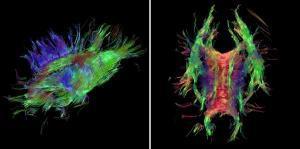
A team of researchers at the Eindhoven University of Technology has developed a software tool that physicians can use to easily study the wiring of the brains of their patients. The tool converts MRI scans using special techniques to three-dimensional images. This now makes it possible to view a total picture of the winding roads and their contacts without having to operate. Researcher Vesna Prčkovska defended her PhD thesis on this subject last week.
To know accurately where the main nerve bundles in the brain are located is of immense importance for neurosurgeons, explains Bart ter Haar Romenij (professor of Biomedical Image Analysis, at the Department of Biomedical Engineering). As an example he cites 'deep brain stimulation', with which vibration seizures in patients with Parkinson's disease can be suppressed. "With this new tool, you can determine exactly where to place the stimulation electrode in the brain.
More at Science Daily
Published on October 31, 2010 02:17
