Deborah Carroll's Blog, page 15
December 23, 2013
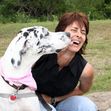
FHO for Westie Going Well Now…
This is the text I got from the owner Saturday, three days later:
Hi, Deborah-
We are seeing good improvement in JJ. He’s standing on his leg at each meal and he has started to use it during our slow walks. He appears to be much happier-caught him smiling today! We haven’t completed all the walks you prescribed yet. Only 2 5 min walks today. He’s keeping still most of the day. If not in his crate, he sleeps on the floor in my office or in our family room. We look forward to seeing you next week.
That’s great news, and we had an hour and a half appointment, in person, to ensure that the caretaker really understood what was going on and my instructions.
It also really, really helps that we have full support from the veterinarian, a young man who does a lot of surgery and with whom I have only worked infrequently. He became familiar with the type of exercise physiology-based rehab I promote and practice through his own life adventures, and he seems to understand and embrace the concepts I talk about with him. He successfully uses a lot of these guidelines without my intervention, and I’m really glad to have been brought in on this case. I think together we’ll make a huge difference!
I’m soon to publish the guidelines for post-FHO surgery, but in the meantime, you will do well to use the same guidelines I have already published for post-knee surgery. Here is a link to the Amazon page with the booklet, currently available in paperback or on Kindle:
http://www.amazon.com/dp/B00EY3D03S
Blessings-
Deborah
Filed under: Hips, Hips Tagged: animal therapy hip surgery, dog pain after surgery, dog rehabilitation after hip surgery, hip surgery, legg calves perthes, westie fho


November 26, 2013
Discount Pricing Available for Orders of 20 or More-
Contact me for discount pricing on bulk orders of my rehab booklets! Clinics are using them to give to clients as discharge instructions and pet-centric businesses are re-selling them outright. Bulk discounts are not currently available if you order from the online source, so let me know your interests and I’ll get pricing to you! Thanks! Blessings
Here is the link to one title on Amazon US, and the titles are available on all the Amazon sites worldwide, so search the title under your country if you have Amazon access:
http://www.amazon.com/dp/B00F7VMJYW
Search under my author profile to find other titles of my rehab guides.
Also should be available worldwide from Barnes & Noble, Bowker distributors, and more! Search and request using the ISBN for any of the titles you want.
Filed under: MEDIA AND PUBLICATIONS Tagged: animal rehabilitation, does my dog need surgery?, exercises for my dog after surgery


November 21, 2013
POST-FHO HOMEWORK SUGGESTIONS FOR CATS
POST-FHO HOMEWORK SUGGESTIONS FOR FELINES
(Femoral Head Ostectomy/Removing the Ball off the Femur at the Hip Joint)
First and foremost: pay attention to the discharge instructions your veterinarian has given you and really try to follow them. These instructions usually include keeping your cat as subdued as possible for at least two weeks, preferably subdued with only controlled activity for 8-12 weeks. I highly recommend you not allow playing, no cat rugby, no no toys, no “I’m still the bossiest kitty” smack-downs from the surgery kitty to the other kitties, etc…and definitely no jumping onto things for 8-12 weeks. Given the opportunity, it is highly likely your cat will escape from you upon arriving home from the hospital and will promptly and speedily dash to some hiding place. It is better to keep your cat in their crate and upon arriving home from the clinic, keep them in a place of your choosing to govern them during this time of healing. I’m pretty sure controlled restriction will work out better than if you end up pulling your cat by the armpits out from under the bed.
During this surgery, there was cutting of muscle and other tissue that will require care and time to heal. Please follow the icing instructions noted on a separate sheet and which are also available on my websites if your cat will allow it.
In physical rehabilitation after FHO we should aim at keeping the “false” joint comfortable after surgery by promoting hip flexion and extension through therapeutic exercises that stimulate leg use, leading to muscle strengthening and avoiding chronic disuse of the operated limb.
The muscle that was cut into during the FHO requires a little over 6 weeks to achieve a normal collagen ratio and will take longer to heal more fully. This should be considered when you think your cat is ready to jump onto high places at 2 weeks after surgery. Don’t let them if you hope for the best outcome from the surgery. On the other hand, the bone that was cut does not require the same care that a fracture repair would; there is no need to be concerned that you will cause further damage to the bone by allowing exercise.
In light of this information I recommend working on some of the rehab activities noted below:
Some cats are accustomed to going on walks, even leash walks, with owners, and if your cat is one of them, then the standard walking homework I write for canines may be followed for cats as well. You may want to try to implement that homework even if you have not previously “walked” your cat. Please use a harness to introduce this walking activity.
During the first two weeks especially, we want your cat to walk and stretch and use their operated leg in a natural, yet controlled manner and with moderate to slow movements. Any walking is fine, i.e., to the litter box, to food and water, but pouncing, jumping and dashing are to be avoided altogether or as much as possible.
If your cat is using the leg fairly well a day or two after surgery, then I encourage you to slowly increase the time of consistent leg use and otherwise start some structured walking at five days after surgery. If your cat will not go for structured walks with you, then another possibility is to use a favorite treat to coax them to walk slowly across the floor. You could hold up the treat at head height and crawl along with your cat to get them to walk along in a continual gait pattern as best possible.
Two to five minutes of this walking a couple of times a day for the first week will be beneficial for the cat and possibly hard on you. This same treat method may be used in conjunction with another person holding a leash on a harness on the cat to introduce the concept of leash walking, which would be easier on you. Some cats will follow a string or feather, etc…pulled slowly across the floor, and this method may be used if your cat will walk sluggishly. However, many cats will wait for distance and then pounce on the string or feather, so use your knowledge of your cat to act as wisely as possible.
The goal is to encourage enough continual, weight-bearing leg use so as to create a callous of scar tissue within the compartment where the top of the femur now rides. Too much activity and/or abrupt, jumping movements could tear up the scar tissue we want and instead create more scarring from the new damage. Eventually, with too much activity, there could be a bulk of scar tissue and increased pain. This doesn’t happen as often in cats as it does in dogs, primarily because they do not weigh as much, and therefore do not put as much pressure on the surgery leg. That extra, harsh, impact pressure is what can cause the top of the femur to tear into the healing area where we’d like to have a callous of scar tissue form. Slow, steady, easy exercise brings the best healing in most cases.
After about five days, and especially if your cat is not using the leg much, then I recommend you speak to your veterinarian about finding some additional pain control medications that will suit your cat. Recovery will improve if your pet feels less pain and is able to use their leg more “normally”, yet gently. Pain medicine helps achieve this, and it seems the medications are needed for an average of 4 weeks for cats after this surgery.
There are a variety of ways to get your cat to stretch out that operated hind leg and any may be utilized as long as the end result is not further injury. I find that with careful restrictions and exercise, along with proper pain medication, cats will usually come around to using their leg as fully as ever without anyone stretching it or forcing movement.
If I see a cat more than 6 weeks out from surgery, and they have plenty of pain medication yet aren’t using the leg in good extension, I will work on exercises and drills that encourage the cat to stretch on their own. Sometimes I get a cat to extend from the floor to a couch for a treat or toy and then I draw them back to the floor again. During the first four weeks this works best if the cat does not end up jumping onto the couch. A stretching drill like this should be done 2-4 times per day with 10 repetitions each time. Please allow your cat to rest and recovery at least 2 hours between exercise sessions.
After three weeks, then more structured play to encourage stretching, leg use and muscle strengthening may be implemented. One example of this is to use a feather or string in the air that your cat will rise onto their hind legs and reach to bat. Two to three minutes of this type of play or twenty repetitions at this time, twice per day is beneficial.
At four weeks, if your cat will walk with you up and down stairs without bounding, it is good to start this exercise. Some cats will follow the owner for continual repetitions. Some cats will need a leash and harness. Where and when possible, a set of five times eight to ten stairs once every other day could be a good workout. Any slow climbing is better than none, and more repetitions in a row serve the muscles better than only one or two stairs here and there.
By three to four weeks, your cat will be wanting to run around more and will function as if they are ready for all the “usual” household activities. I still prefer to avoid very harsh movements at this time so that cats don’t disrupt the good scar tissue that has formed. I tell people that the tissue we want is very much like what you get when you ride a bike a lot. If you have not ridden in a while and you go out for a longer ride, the bones at your seat will likely feel like they hurt the next day when you sit in a hard chair. People who frequently ride have scar tissue that operates as padding between bone and tissue. After a couple of riding sessions, the appropriate scar tissue forms and it is no longer painful to sit. This is very similar to the type of tissue I want to see your cat form after an FHO; they need a slow build-up of scar tissue to cushion between the femur and the muscle, and while it is forming due to friction from consistent leg use, I don’t want to tear it or otherwise disrupt it with harsh movements. That could lead to formation of more bulky scar tissue which makes it harder for the leg to move and sometimes causes nerve pain. Similarly, we don’t want to allow the animal to not use the leg, because scar tissue will form that will bind the leg into a place of diminished function and it will always then hurt to do some favorite activities in the future. Not too much, not too little.
If your pet is not using the operated leg after week 1, then I recommend calling your vet or me for rehab intervention and to get them started on beneficial exercise and pain medications. If you follow the exercise prescription well and after week 4 would like advanced exercises, then a rehab consult is necessary. I have some separate recommendations for canine FHO’s, so feel free to contact your vet for a copy of that if you need it.
And if your cat does end up hiding under the bed when you get home because you felt sorry for them and let them out of their crate, don’t pull them out from under the bed by the armpits J I do recommend that you shut the doors to the bedroom, closet, and bathroom, though, so that when they do come out from under the bed, you will have a better chance of collecting them and getting them back into a crate.
©2007 Rehabilitation and Conditioning for Animals
Deborah Carroll CCRP, CSCS
Filed under: Hips Tagged: cat hip surgery, cat rehab after surgery, exercises for my cat after surgery, FHO, rehabilitation for cats, therapy for cat


November 19, 2013
Rehab Booklets Now Available on Amazon Australia!
Here is the link to Guidelines for Home Rehabilitation of Your Dog: Instead of Surgery for Torn Knee Ligament: The First Four Weeks, Basic Edition:
https://www.amazon.com.au/dp/B00F7VMJYW
which is also available in paperback in a variety of markets,
and here is the link to After Surgery for Torn Knee Ligament, The First Four Weeks, Basic Edition
https://www.amazon.com.au/dp/B00EY3D03S
I’m excited about this news, because I have a lot of activity on this site from Australia…but I’m first a fan of paper books and small booksellers, so I’m really interested to get the paperback versions in Australia!! Working on it, and I’ve registered with distributors…wish I had clones so I could follow up every detail “perfectly” and be able to find the “best” option for any and every one to get this information, if they want it, in the “best” format possible.
In the meantime, since the paperbacks became available last month, clinics are ordering in bulk to pass out copies to clients when the need arises, and that’s a good thing 
Thanks
Filed under: Knees (Stifles, Patellas) Tagged: animal rehabilitation, dog rehabilitation australia, dog therapy, dog torn knee ligament


November 9, 2013
Knees (Stifles, Patellas) – 5 Articles
“Surgery may not always be the best first course of action. A physical therapist, in many cases, can help patients avoid the often unnecessary risks and expenses of surgery. This study should help change practice in the management of symptomatic meniscal tears in patients with knee osteoarthritis.” Mar. 21, 2013 — A New England Journal of Medicine (NEJM) study showing that physical therapy is just as effective as surgery in patients with meniscal tears and arthritis of the knee should encourage many health care providers to reconsider their practices in the management of this common injury, according to the American Physical Therapy Association (APTA).
The study, published March 19, showed no significant differences in functional improvement after 6 months between patients who underwent surgery with postoperative physical therapy and those who received standardized physical therapy alone.
“This study demonstrates what physical therapists have long known,” explained APTA President Paul A. Rockar Jr, PT, DPT, MS. “Surgery may not always be the best first course of action. A physical therapist, in many cases, can help patients avoid the often unnecessary risks and expenses of surgery. This study should help change practice in the management of symptomatic meniscal tears in patients with knee osteoarthritis.” According to lead physical therapist for the trial and American Physical Therapy Association (APTA) member Clare Safran-Norton, PT, PhD, OCS, “our findings suggest that a course of physical therapy in this patient population may be a good first choice since there were no group differences at 6 months and 12 months in this trial. These findings should help surgeons, physicians, physical therapists, and patients in decision-making regarding their treatment options.” Researchers at 7 major universities and orthopedic surgery centers around the country studied 351 patients aged 45 years or older who had a meniscal tear and mild-to-moderate osteoarthritis of the knee. Patients were randomly assigned to groups who received either surgery and postoperative physical therapy or standardized physical therapy. Within 6-12 months, patients who had physical therapy alone showed similar improvement in functional status and pain as those who had undergone arthroscopic partial meniscectomy surgery. Patients who were given standardized physical therapy — individualized treatment and a progressive home exercise program — had the option of “crossing over” to surgery if substantial improvements were not achieved. Thirty percent of patients crossed over to surgery during the first 6 months. At 12 months these patients reported similar outcomes as those who initially had surgery. Seventy percent of patients remained with standardized physical therapy. According to an accompanying editorial in NEJM,”millions of people are being exposed to potential risks associated with a treatment [surgery] that may or may not offer specific benefit, and the costs are substantial.” Physical therapist and APTA member Mary Ann Wilmarth, PT, DPT, MS, OCS, MTC, Cert MDT, chief of physical therapy at Harvard University, said, “Physical therapists are experts in improving mobility and restoring motion. The individualized treatment approach is very important in the early phases of rehabilitation in order to achieve desired functional outcomes and avoid setbacks or complications.”
Story Source: The above story is reprinted from materials provided by American Physical Therapy Association. Note: Materials may be edited for content and length. For further information, please contact the source cited above.
Journal Reference:
Jeffrey N. Katz, Robert H. Brophy, Christine E. Chaisson, Leigh de Chaves, Brian J. Cole, Diane L. Dahm, Laurel A. Donnell-Fink, Ali Guermazi, Amanda K. Haas, Morgan H. Jones, Bruce A. Levy, Lisa A. Mandl, Scott D. Martin, Robert G. Marx, Anthony Miniaci, Matthew J. Matava, Joseph Palmisano, Emily K. Reinke, Brian E. Richardson, Benjamin N. Rome, Clare E. Safran-Norton, Debra J. Skoniecki, Daniel H. Solomon, Matthew V. Smith, Kurt P. Spindler, Michael J. Stuart, John Wright, Rick W. Wright, Elena Losina. Surgery versus Physical Therapy for a Meniscal Tear and Osteoarthritis. New England Journal of Medicine, 2013; : 130318220107009 DOI:10.1056/NEJMoa1301408
Note: If no author is given, the source is cited instead.
Here is a second report of the same issue:
Medscape Medical News from the:
American Academy of Orthopaedic Surgeons (AAOS) 2013 Annual Meeting
This coverage is not sanctioned by, nor a part of, the American Academy of Orthopaedic Surgeons.
Medscape Medical News > Conference News
Physical Therapy as Effective as Surgery for Meniscal Tear
Medscape Medical News from the: American Academy of Orthopaedic Surgeons (AAOS) 2013 Annual Meeting Physical Therapy as Effective as Surgery for Meniscal Tear Kathleen Louden Mar 20, 2013 CHICAGO, Illinois — Patients with knee osteoarthritis and a meniscal tear who received physical therapy without surgery had good functional improvement 6 months later, and outcomes did not differ significantly from patients who underwent arthroscopic partial meniscectomy, a new clinical trial shows. In the Meniscal Tear in Osteoarthritis Research (METEOR) trial, both groups of patients improved substantially in function and pain. This finding, presented here at the American Academy of Orthopaedic Surgeons 2013 Annual Meeting and published online simultaneously in the New England Journal of Medicine, provides “considerable reassurance regarding an initial nonoperative strategy,” the investigators report. Patients with a meniscal tear and osteoarthritis pose a treatment challenge because it is not clear which condition is causing their symptoms,” principal investigator Jeffrey Katz, MD, from Brigham and Women’s Hospital in Boston, Massachusetts, told Medscape Medical News. “These data suggest that there are 2 reasonable pathways for patients with knee arthritis and meniscal tear,” Dr. Katz explained. “We hope physicians will use these data to help patients understand their choices.” In an accompanying editorial, clinical epidemiologist Rachelle Buchbinder, PhD, from the Monash University School of Public Health and Preventive Medicine in Victoria, Australia, said that “these results should change practice. Currently, millions of people are being exposed to potential risks associated with a [surgical] treatment that may or may not offer specific benefit, and the costs are substantial.” These results should change practice. The METEOR trial enrolled 351 patients from 7 medical centers in the United States. Eligible patients were older than 45 years, had osteoarthritic cartilage change documented with magnetic resonance imaging, and had at least 1 symptom of meniscal tear, such as knee clicking or giving way, that lasted at least 1 month despite drug treatment, physical therapy, or limited activity. In this intent-to-treat analysis, investigators randomly assigned 174 patients to arthroscopic partial meniscectomy plus postoperative physical therapy and 177 to physical therapy alone. The physical therapy in both regimens was a standardized 3-stage program that allowed patients to advance to the next intensity level at their own pace, Dr. Katz explained. The program involved 1 or 2 sessions a week for about 6 weeks and home exercises. The average number of physical therapy visits was 7 in the surgery group and 8 in the nonsurgery group. Investigators evaluated patients 6 and 12 months after randomization. The primary outcome was the between-group difference in change in physical function score from baseline to 6 months, assessed using the Western Ontario and McMaster Universities Osteoarthritis Index (WOMAC). At baseline, demographic characteristics and WOMAC physical function scores were similar in the 2 groups. At 6 months, improvement in the WOMAC function score was comparable in the 2 groups. The mean between-group difference of 2.4 points was not statistically significant after analysis of covariance. There was also no significant difference between groups in pain improvement or frequency of adverse events. METEOR: Mean Improvement in Osteoarthritis Index at 6 Months Treatment Group Mean Improvement (Points) 95% Confidence Interval Surgery plus physical therapy 20.9 17.9–23.9 Physical therapy 18.5 15.6–21.5 There was 1 death in each group, and 8 patients in the nonsurgery group and 13 in the surgery group withdrew in the first 6 months of the study. Patients in the nonsurgery group were allowed to cross over to the surgical group at any time. Within 6 months, 30% of patients did so. “They were not doing very well,” Dr. Katz said. His team is still analyzing the reasons these patients did not benefit from intensive physical therapy. The 12-month results were similar to the 6-month results. In addition, by 12 months, outcomes for the crossover patients and for those in the original surgery group were similar. Meeting delegate John Mays, MD, an orthopaedic surgeon practicing in Bossier City, Louisiana, who was asked by Medscape Medical News to comment on the findings, said most patients don’t choose physical therapy. “In the real world, most people want a quick fix” and choose surgery, he noted. Dr. Mays said he would have liked to have seen a group of patients who underwent surgery but did not receive postoperative physical therapy. He explained that his patients with osteoarthritis and meniscal tear rarely get physical therapy after arthroscopic meniscectomy; they most often do home-based exercises. He added that “most insurance plans have limits on the number of physical therapy sessions they allow.” This study is funded by the National Institute of Arthritis and Musculoskeletal and Skin Diseases. Dr. Katz, Dr. Buchbinder, and Dr. Mays have disclosed no relevant financial relationships. N Engl J Med. Published online March 19, 2013. Abstract, Editorial American Academy of Orthopaedic Surgeons (AAOS) 2013 Annual Meeting: Abstract SE67. Presented March 19, 2013.
More Than Half of All ACL Reconstructions Could Be Avoided, Five-Year Follow-Up Study Shows
(From RehabDeb: This report is from human medical research, however animal studies are currently being conducted at Colorado State University. When I began animal rehab in 2005, I developed some protocol for people to use to benefit their animals if they did not want surgery for their pet, even though I was working at the time in a surgery specialty hospital. When I began independent practice in 2007, I took years of accumulated research, experience, and knowledge and created some simple functional exercise and drill protocol that has benefited hundreds of my canine patients whose people opted to not pursue surgery. That protocol and some other papers citing surgery text recommendations may be found elsewhere on this site-see the index to the right. In every case where my protocol has been followed (and there are no extenuating circumstances), the pets have stabilized the joint with muscle and scar tissue, and they have functioned very well. This work is all done in the home environment with no dependence on specialized equipment…no need when we are drawing from centuries of known exercise physiology and dynamic principles of body function. Blessings-)
Jan. 30, 2013 — In the summer of 2010, researchers from Lund University in Sweden reported that 60 per cent of all anterior cruciate ligament (ACL) reconstructions could be avoided in favour of rehabilitation. The results made waves around the world, and were met with concerns that the results would not hold up in the long term. Now the researchers have published a follow-up study that confirms the results from 2010 and also show that the risk of osteoarthritis and meniscal surgery is no higher for those treated with physiotherapy alone.
“We have continued with our study and for the first time are able to present a five-year follow-up on the need for and results of ACL surgery as compared with physiotherapy. The findings have been published in the British Medical Journal and are basically unchanged from 2010. This will no doubt surprise many people, as we have not seen any difference in the incidence of osteoarthritis,” says Richard Frobell, one of the researchers behind the study, who is an associate professor at Lund University and a clinician at the orthopaedic department, Helsingborg Hospital.
Richard Frobell explains that the research group’s results from 2010, which were published in the New England Journal of Medicine, caused a stir and questions were raised as to whether it was possible to say that an operation would not be needed in the long term.
Half of the patients who were randomly assigned not to undergo reconstructive surgery have had an operation in the five years since, after experiencing symptoms of instability.
“In this study, there was no increased risk of osteoarthritis or meniscal surgery if the ACL injury was treated with physiotherapy alone compared with if it was treated with surgery. Neither was there any difference in patients’ experiences of function, activity level, quality of life, pain, symptoms or general health,” says Richard Frobell.
“The new report shows that there was no difference in any outcome between those who were operated on straight away, those who were operated on later and those who did not have an operation at all. The message to the medical experts who are treating young, active patients with ACL injuries is that it may be better to start by considering rehabilitation rather than operating straight away.”
In Sweden, over 5 000 people every year suffer an anterior cruciate ligament injury — mainly young people involved in sport. There are different schools of treatment and Sweden stands out with treatment that is in line with the results of the study.
“On an international front, almost all of those with ACL injuries are operated on. In Sweden, just over half are operated on, but in southern Sweden we have been working for many years to use advanced rehabilitation training as the first method of treatment. Our research so far has confirmed that we are right in not choosing to operate on these injuries immediately. Longer-term follow-up is important, however, if we are to look more closely at the development of osteoarthritis in particular,” says Richard Frobell.
The research group, whose study is called KANON, Knee ACL NON-operative versus operative treatment, is now moving on to the next stage. This year, the third part of the study will begin, following up the patients ten years after anterior cruciate ligament injury.
Richard Frobell has also entered into a collaboration with researchers at the School of Economics and Management at Lund University to evaluate the health economics aspects of different treatment methods for ACL injury.
Journal References:
R. B. Frobell, H. P. Roos, E. M. Roos, F. W. Roemer, J. Ranstam, L. S. Lohmander. Treatment for acute anterior cruciate ligament tear: five year outcome of randomised trial. BMJ, 2013; 346 (jan24 1): f232 DOI:10.1136/bmj.f232
Richard B. Frobell, Ewa M. Roos, Harald P. Roos, Jonas Ranstam, L. Stefan Lohmander. A Randomized Trial of Treatment for Acute Anterior Cruciate Ligament Tears.New England Journal of Medicine, 2010; 363 (4): 331 DOI:10.1056/NEJMoa0907797
From ScienceDaily
Stifle (Knee) Ligament Ruptures (Torn ACL, CCL) Information Overview
(This post contains information about ligaments and cites surgery recommendations and some rehab possibilities. The homework protocol I have written for use after surgery and/or instead of surgery and which has been used successfully for years is now available in book form, and here are the links: http://www.amazon.com/dp/B00F7VMJYW and http://www.amazon.com/dp/B00EY3D03S)
Ligaments are dense connective tissue structures consisting of fibroblasts, water, collagen, proteoglycans, fibronectin, and elastin that connect two or more bones (1, 2). Currently, a great deal of information remains unanswered regarding timing of ligamentous healing, especially with respect to postoperative mobilization techniques (graft, suture, TPLO, etc…). This is because ligaments heal differently depending on the location. For example, the healing potential of the medial collateral ligament of the stifle is very good, but the cranial cruciate ligament, which has received the most investigation, demonstrates virtually no healing response following injury (2). Within hours of injury, the defect is filled with an organized hematoma and the surrounding tissue becomes edematous from perivascular leakage of fluid. Monocytes and macrophages are found in the wound by 24 hours and respond by cleaning up the site and transitioning to the next phase.
This acute injury phase lasts approximately 48-72 hours (2). In other words, the knee will swell, sometimes only a little, inside the joint, thus making the bony parts thicker or wider, expanded due to fluid accumulation. Other times there is swelling in the soft tissue as well. It is during this acute phase that the use of ice is recommended, 1-6 times per day, for 20 minutes each application, on average, depending on fur density and type of ice used. I have a separate paper with icing recommendations on this site, under “Homework”. The method of delivery most recommended yet one of the least effective is frozen veggies, so check out the other options noted in the other paper. The use of heat on an acute injury is not recommended and will likely be destructive to the natural healing process. Again, do not use heat on the knee injury at this time. During this time and throughout the healing process the use of low-level laser therapy is also warranted.
More research in recent years has shown that stopping the inflammatory process, a process that is a natural part of healing, is not a good idea much of the time for this type of injury. If the body is allowed to go through the inflammatory process, especially if there are pain medications like Tramadol available, then healing may occur faster. Ice and nsaids, non-steroidal anti-inflammatories, work against inflammation, and nsaids also work against healing. The main idea is to lower the level of pain and to encourage healing, so use the best tools and information you have available.
In many cases, loss of ligamentous support invariably leads to progressive osteoarthritis, such as in cranial cruciate ligament (ACL) ruptures. Please note that the arthritic process began when the first disruptions occurred in the joint, when damage first occurred and then when tearing first began. A ligament usually will tear for some time prior to total rupture. A ligament rupture is not a matter of life and death. Many people come to me relaying that they have felt rushed into or forced toward surgery for this condition in their dog. In contrast, I had one client who is a human medical doctor state, “I wish we could get people off of the surgery idea…we don’t even rush every human athlete into surgery, much less every person in general.”
Slatter’s Textbook of Small Animal Surgery, 2nd Edition states that small dogs often do well without surgical intervention, and that based on particular studies, “it is prudent to wait for at least 6 to 8 weeks before recommending surgery for small dogs. These dogs are older at diagnosis and are often obese with concurrent medical problems. Small dogs that are lame for 6 weeks after diagnosis and show no improvement often have meniscal tears and are operated on for meniscectomy and joint stabilization.” (pg.1832) Your veterinarian or I may help you evaluate whether or not your dog has a meniscal tear.
Additionally, I have successfully used basic and advanced functional rehab protocol I developed based on principles of athletic training to address joint instability and muscle atrophy that occurs along with torn and ruptured knee ligaments in large dogs. Some positive feedback from veterinarians and owners is cited on my websites and in separate blog posts regarding this exercise protocol.
Excessive exercise during periods of acute joint inflammation may be detrimental to articular cartilage, and immobilization may be protective during acute bouts of inflammatory joint disease. (4) Joint inflammation will occur with greater stresses that are placed on the joint in the presence of ligament damage. If surgery is not opted, then for a period of time, depending on the severity of the injury, short, controlled leash walks and restricted activity along with mandatory rest are indicated during the first phase of acute injury. Even if surgery is opted, the recovery time and exercise protocol are virtually the same.
There were no written protocol I found within veterinary medicine addressing specific exercise protocol and return to function when I began practicing functional rehab in veterinary medicine in 2005. I subsequently began writing protocol based on how similar human injuries are managed and treated, from athletes to sedentary individuals.
Given that we have discussed loss of support and inflammation of ligament and joint, it would follow that muscle atrophy would be another complication to address. Muscle atrophy will occur whether or not surgery is performed, and rehab interventions are proven to aid in gaining strength and muscle tone in the affected limb. Muscle atrophy has been occurring during the whole time the pet has been injured because the injury will have produced pain and instability, even if mild at first, and that will have encouraged disuse and, therefore, atrophy.
The degree of quadriceps muscle atrophy present before surgery for CCL rupture seems to correlate significantly with the degree of cartilage fibrillation, indicating a relationship with the severity of the condition. In studies, muscle mass improved 7 and 13 months after surgery, but significant residual muscle atrophy remained in many dogs even after 1 year. I usually see muscle atrophy reversed in much less time when owners have followed recommended protocol.
A specific exercise program with changes in protocol as time goes on will indeed build muscle and will usually cause hypertroph better than surgical repair alone or pain medication alone, based on observations. Research citations to validate this foundational truth may be found elsewhere in this blog or in a bazillion places online. Try http://www.nsca-lift.org for foundations in strength training if you have further interest in this specialty.
Outside the scope of this writing is the argument as to whether a natural course of events follows evolution or deterioration without intervention; either way it is the primary purpose of rehabilitation interventions to improve upon what natural abilities would theoretically otherwise be realized. Whether or not an animal will do well on its own without intervention is inconsequential when the overwhelming benefits of rehabilitation intervention are considered. In light of this, rehabilitation treatment is indicated whether or not ligament repair surgery is performed.
For non-surgical patients, rehab treatment may consist of conservative exercise that increases in difficulty as healing progresses and of therapies such as massage, low-level laser, ice, ultrasound, nutraceuticals, and weight control plans. I find assisted, forced specific range of motion exercises to be unnecessary in a companion animal that is functional, one that is able to move their limbs on their own.
For non-surgical patients, building muscle and supporting tissue will be important as well as maintaining protective interventions for affected joints, i.e., the use of therapies mentioned and maintaining dosing supplements and/or pharmaceuticals proven to aid. Nutrition supplement support, or nutraceuticals, proven to aid include glucosamine/chondroitin/MSM (all work better together) and fish oil. There are others. It is also outside the scope of this writing to argue or discuss the benefits of the nutraceuticals mentioned.
A qualified rehabilitation practitioner in collaboration with the treating veterinarian should be able to design a basic appropriate plan of action to meet your and your pet’s needs. It is within the scope of this paper to briefly and generally give information regarding ligament damage and specifically cruciate ligament damage. The conclusion is that if this information generates more questions, then answers should be sought from a qualified animal health care professional. Additionally, please see my separate post regarding homework recommendations for torn knee ligaments. Thank you!
References: 1. Fowler D: Principles of wound healing. In Harari J, editor: Surgical complications and wound healing in the small animal practice, Philadelphia, 1993, WB Saunders. 2. Frank C et al: Normal ligament: structure, function, and composition. In Woo S, Buckwalter J, editors: Injury and repair of the musculoskeletal soft tissues, Park Ridge, Illinois, 1991, American Academy of Orthopedic Surgeons Symposium. 3. Moore KW, Read RA: Rupture of the cranial cruciate ligament in dogs. II. Diagnosis and management, Compendium of Continuing Education Pract Vet 18:381391, 405, 1996 4. Agudelo CA, Schumacher HR, Phelps P: Effect of exercise on urate crystal-induced inflammation in canine joints, Arthritis Rheum 15:609-616, 1972
Copyright 2007, Deborah Carroll
Quality of Life of Obese Dogs Improves When They Lose Weight
This is actually recent research that was done in the UK, where they estimate 1/3 of the dog population is obese. Study conducted by Waltham/Royal Canin. I wouldn’t think we needed research to prompt us on this, however human nature proves we do! For those of you who need research to tell you that your dog will have a longer, happier life (same goes for humans…) if they drop the extra fat, here it is! Wheeeeeee!
The results showed that the quality of life improved in the dogs that had successfully lost weight, in particular vitality scores increased and the score for emotional disturbance and pain decreased. Moreover, the more body fat that the dog lost, the greater the improvement in vitality.
…and, interestingly, the study notes this: “The research also found that dogs that failed to complete their weight loss programme had worse quality of life at the outset than those successfully losing weight, most notably worse vitality and greater emotional disturbance.” …sort of as if the dogs failed the program and not that the owners were part and parcel. lol…the dogs didn’t fail to complete the program. And their finding here denotes the close connection and issues to be explored within the human/animal psychology bond; it works both ways-to the positive and to negative effect. The failed dogs notably had ‘worse quality of life at the outset” than the ones who ended up succeeding, and most compromised were their vitality and emotional status. We definitely pass our moods, demeanor, and worry onto our animals. Breathe peacefully with your pets
The “HOW TO” is up to me to help you accomplish, usually in conjunction with your vet.
I have over 30 years experience in program design and nutrition, so I am well qualified to help with lifestyle changes and subtle or great control measures. People are usually able to follow my plans because I work with them to determine how they operate best, whether with large changes or baby steps or in-between. Blessings-Feb. 21, 2012 — Researchers at the University of Liverpool have found that overweight dogs that lose weight have an improved quality of life compared to those that don’t.
A study of 50 overweight dogs, comprising a mix of breeds and genders was undertaken by scientists at the University in collaboration with the University of Glasgow, Royal Canin and the WALTHAM Centre for Pet Nutrition.
Owners completed a questionnaire to determine the health-related quality of life of their dog prior to weight loss. A follow-up questionnaire was completed by the owners of 30 dogs that successfully completed the weight loss programme, enabling changes in quality of life to be assessed. A range of life quality factors were scored, including vitality, emotional disturbance and pain. The quality of life of dogs which succeeded with their weight loss programme was also compared with those dogs that failed to lose weight successfully.
The results showed that the quality of life improved in the dogs that had successfully lost weight, in particular vitality scores increased and the score for emotional disturbance and pain decreased. Moreover, the more body fat that the dog lost, the greater the improvement in vitality.
The research also found that dogs that failed to complete their weight loss programme had worse quality of life at the outset than those successfully losing weight, most notably worse vitality and greater emotional disturbance.
Dr Alex German, Director of the Royal Canin Weight Management Clinic at the University, said: “Obesity is a risk for many dogs, affecting not only their health but also their quality of life. This research indicates that weight loss can play an important role in keeping your dog both healthy and happy.”
Dr Penelope Morris, from the WALTHAM Centre for Pet Nutrition, added: “Strategies for combating obesity and keeping dogs fit and healthy include portion control, increased exercise and diets specifically formulated for overweight pets.”
Established in 2004, the Royal Canin Weight Management Clinic at the University’s Small Animal Hospital UK’s is the world’s first animal weight management referral clinic and was set up to help tackle and prevent weight problems in animals such as dogs and cats.
Veterinary surgeons from any general practice in the UK can refer overweight animals to the clinic. The patients receive a thorough medical examination, and are then given a specific dietary plan and exercise regime to follow over several weeks.
Taken from ScienceDaily
Filed under: RESEARCH CITATIONS (NOT EXHAUSTIVE :))


Cognitive and Neurological – 5 Articles
ScienceDaily (June 1, 2012) — Exercise helps to alleviate pain related to nerve damage (neuropathic pain) by reducing levels of certain inflammation-promoting factors, suggests an experimental study in the June issue of Anesthesia & Analgesia, official journal of the International Anesthesia Research Society (IARS).
The results support exercise as a potentially useful nondrug treatment for neuropathic pain, and suggest that it may work by reducing inflammation-promoting substances called cytokines. The lead author was Yu-Wen Chen, PhD, of China Medical University, Taichung, Taiwan.
Exercise Reduces Nerve Pain and Cytokine Expression in Rats Neuropathic pain is a common and difficult-to-treat type of pain caused by nerve damage, seen in patients with trauma, diabetes, and other conditions. Phantom limb pain after amputation is an example of neuropathic pain.
Dr Chen and colleagues examined the effects of exercise on neuropathic pain induced by sciatic nerve injury in rats. After nerve injury, some animals performed progressive exercise — either swimming or treadmill running — over a few weeks. The researchers assessed the effects of exercise on neuropathic pain severity by monitoring observable pain behaviors.
The results suggested significant reductions in neuropathic pain in rats assigned to swimming or treadmill running. Exercise reduced abnormal responses to temperature and pressure — both characteristic of neuropathic pain.
Exercise also led to reduced expression of inflammation-promoting cytokines in sciatic nerve tissue — specifically, tumor necrosis factor-alpha and interleukin-1-beta. That was consistent with previous studies suggesting that inflammation and pro-inflammatory cytokines play a role in the development of neuropathic pain in response to nerve injury.
Exercise also led to increased expression of a protein, called heat shock protein-27, which may have contributed to the reductions in cytokine expression.
Neuropathic pain causes burning pain and numbness that is not controlled by conventional pain medications. Antidepressant and antiepileptic drugs may be helpful, but have significant side effects. Exercise is commonly recommended for patients with various types of chronic pain, but there are conflicting data as to whether it is helpful in neuropathic pain.
The new results support the benefits of exercise in reducing neuropathic pain, though not eliminating it completely. In the experiments, exercise reduced abnormal pain responses by 30 to 50 percent.
The study also adds new evidence that inflammation contributes to the development of neuropathic pain, including the possible roles of pro-inflammatory cytokines. The results provide support for exercise as a helpful, nondrug therapy for neuropathic pain — potentially reducing the need for medications and resulting side effects.
Practice Makes the Brain’s Motor Cortex More Efficient
**Repetitive motion drills are key to rehab protocol I design for animals with neurological defecits—so keep on keeping on if you are doing that, too! Blessings-RehabDeb
Aug. 4, 2013 — Not only does practice make perfect, it also makes for more efficient generation of neuronal activity in the primary motor cortex, the area of the brain that plans and executes movement, according to researchers from the University of Pittsburgh School of Medicine. Their findings, published online today in Nature Neuroscience, showed that practice leads to decreased metabolic activity for internally generated movements, but not for visually guided motor tasks, and suggest the motor cortex is “plastic” and a potential site for the storage of motor skills.
The hand area of the primary motor cortex is known to be larger among professional pianists than in amateur ones. This observation has suggested that extensive practice and the development of expert performance induces changes in the primary motor cortex, said senior investigator Peter L. Strick, Ph.D., Distinguished Professor and chair, Department of Neurobiology, Pitt School of Medicine.
Prior imaging studies have shown that markers of synaptic activity, meaning the input signals to neurons, decrease in the primary motor cortex as repeated actions become routine and an individual develops expertise at a motor skill. The researchers found that markers of synaptic activity also display a marked decrease in monkeys trained to perform sequences of movements that are guided from memory — an internally generated task — rather than from vision. They wondered whether the change in synaptic activity indicated that neuron firing also declined. To examine this issue they recorded neuron activity and sampled metabolic activity, a measure of synaptic activity in the same animals.
All the monkeys were trained on two tasks and were rewarded when they reached out to touch an object in front of them. In the visually guided task, a visual target showed the monkeys where to reach and the end point was randomly switched from trial to trial. In the internally generated task the monkeys were trained to perform short sequences of movements without visual cues. They practiced the sequences until they achieved a level of skill comparable to an expert typist.
The researchers found neuron activity was comparable between monkeys that performed visually guided and internally generated tasks. However, metabolic activity was high for the visually guided task, but only modest during the internally generated task.
“This tells us that practicing a skilled movement and the development of expertise leads to more efficient generation of neuron activity in the primary motor cortex to produce the movement. The increase in efficiency could be created by a number of factors such as more effective synapses, greater synchrony in inputs and more finely tuned inputs,” Dr. Strick noted. “What is really important is that our results indicate that practice changes the primary motor cortex so that it can become an important substrate for the storage of motor skills. Thus, the motor cortex is adaptable, or plastic.
Exercise for Parkinson’s Patients
(With adept application, many human interventions may be crossed over to non-human animal functional rehab  )
)
Becky G. Farley, PhD, PT, MS, knows that exercise is about more than fitness-it is a physiological tool that encourages the body’s own endogenous brain repair mechanisms.
“Exercise promotes brain health and, thereby, may protect the remaining ‘viable’ dopamine neurons, called neuroprotection,” she said.
It also optimizes brain function through activity-dependent plasticity mechanisms that can restore function to damaged pathways, normalize interference from inefficient signaling and increase reliance on undamaged systems.
In July 2010, Dr. Farley founded NeuroFit NetWorks, a non-profit program dedicated to developing and expanding access to research-based exercise programming that is proactive, optimizes brain health/function, and changes the lives of individuals living with a neurodegenerative disease.
Exercise, she said, may at the very least slow motor deterioration in patients with Parkinson’s disease, and, if started early enough, may be able to modify disease progression.
Research, Advocacy & Education
While at the University of Arizona, Dept. of Physiology, Dr. Farley researched muscle activation mechanisms that underlie one of the primary symptoms in patients with Parkinson’s disease. This research led to the development of an exercise program called LSVT BIG™ to target bradykinesia (a slowness of movement) and an NIH-funded randomized clinical trial.
After training more than 5,000 physical and occupational therapists around the world in how to instruct people with Parkinson’s disease in LSVT BIG™ methods, Dr. Farley realized that basic and clinical science research about exercise and Parkinson’s disease is not being translated to real-world application.
“For example, continuous access to proactive neuroplasticity-principled programs that have been shown in animals to slow disease progression.are not available,” she explained. “As it stands now, patients with Parkinson’s disease rarely go to therapy, and when they do, it is usually only after they lose function and start to fall. And those that do go are rarely seen by PD-exercise experts.”
Even if they do find therapists that understand Parkinson’s disease, most patients are not implementing approaches that adhere to the principles of practice that are required to promote learning and plasticity. Instead, outdated guidelines are implemented that promote using strategies or working on secondary impairments like weakness and flexibility.
“No doubt these things help make life better for patients with Parkinson’s disease, but they don’t target the problems that got them to that level of disability. So, ultimately nothing is changed,” said Dr. Farley.
NeuroFit NetWorks plans to change that. The non-profit is advocating for translation of this research now. They have started a national Parkinson Exercise Revolution to help people get better and stay better with exercise. Dr. Farley is conducting training workshops nationally for therapists and fitness professionals to become Parkinson’s disease exercise experts and to work together in their local communities to implement early intervention and continuous access to PD-specific exercise for life.
A model neuro-fitness center in Tucson, AZ, is currently being developed to show how rehab can be accessible and integrated with community fitness programming to optimize learning and function.
“This is the infrastructure that we think offers promise to delay disease onset, slow disease progression, restore motor function, and increase longevity and quality of life for people with neurodegenerative or neurological conditions, and for those individuals at risk for these conditions,” Dr. Farley noted.
It will take advocacy; education of the medical, fitness community and lay public; and working with health care systems to change existing paradigms, demonstrate health care cost savings, and document slowing of motor deterioration.
Research-Based Exercise Programs
All exercise programs at NeuroFit NetWorks are implemented within the context of the Exercise4BrainChange™ (E4BC) model that requires therapists and fitness professionals to incorporate instructions, feedback, and research techniques that promote four essential learning constructs: prepare, activate, reflect, motivate.
These constructs can accommodate multiple exercise approaches and be customized to address a disease’s specific physical/cognitive/emotional deficits. The depth of the content can be modified to allow for a system of communication with a similar language across a variety of therapeutic disciplines and exercise professionals. This is the first time that these essential principles have been described in a manner that can help clinicians implement these concepts immediately with their patients.
“We believe to effect disease modification, proactive models of health care continuums must embrace research-based exercise approaches that are guided by the essential principles of learning and neuroplasticity,” said Dr. Farley. “We call these essential elements “Exercise4BrainChange™ principles” and we have developed a model that integrates all these elements in a way that can promote optimal brain function and skill acquisition.”
Every staff member has undergone training to learn about Parkinson’ disease and exercise and how to modify their instruction/programming to make it PD-specific and optimize the potential for learning and plasticity.
The following programs are offered at the Parkinson Wellness Recovery (PWR!) Gym:
Rehabilitation – 1:1 Exercise4BrainChange. This is where an individual comes to the gym to get a ‘PWR! PLAN.’ They work one-on-one with a therapist that is a PD-exercise expert who educates them about the research on exercise and helps them develop a proactive PD-specific “Use It Or Lose It” program that includes ongoing coaching and tune-ups for life.
Other 1:1 rehabilitation treatments address and target specific problems, such as freezing or postural instability, to “use it and improve it.”
“We integrate research techniques to target bradykinesia/rigidity/coordination/postural instability/posture, such as the training of activated large amplitude whole-body movements; focused practice on axial rotation and extension exercises; paced, rhythmical movements to augmented sensory proprioceptive feedback; rhythmical; treadmill activities for endurance, coordination for gait and balance, and more,” Dr. Farley explained. Clients are progressively challenged to work harder than then self-select. Language and cognitive activities are integrated to increase difficulty, and emotional deficits are targeted through empowerment/education/affirmations to retrain their emotional brain about what they CAN do.
Community – Group E4BC Programming. This program offers PWR! MOVES; PWR! Circuit and specialty classes (agility, strengthening, stretching) that target PD-specific symptoms; and cardio programs for brain health and to prepare the brain to learn! (mobilize neurotransmitters, cell survival and growth factors, boost the immune system, reduce inflammation).
Other enrichment programming to optimize brain health and restoration includes nutrition, stress reduction, brain fitness, and other social or general group exercise activities (drumming, dance, tai chi, etc.).
Retraining the Brain
It is not enough to simply exercise when one has a neurodegenerative disease, noted Dr. Farley.
“It is important to target the anticipated and existing problems with intensive, repetitive practice while receiving certain types of feedback to help you learn and pay attention to critical aspects of the practice,” she explained.
It is also vital that a person is ready to learn and able to manage stress, anxiety and fear to optimize the conditions for learning. That’s why NeuroFit NetWorks has specially trained therapists and fitness professionals that understand how to implement programming to optimize brain health and function for people with different types of conditions.
“Our movements and ability to learn new skills requires active engagement. So, dysfunction in emotional or cognitive systems that interferes with planning, intrinsic motivation, self-monitoring, confidence, etc., interferes with learning,” explained Dr. Farley.
The cognitive deficits in Parkinson’s disease are in the area of executive functioning and attention. These cognitive components are essential for the planning and production of complex whole-body movements; ability to adapt movements/postures to changes in the environment, and automaticity (ability to divide or focus attention for multitasking).
In addition, the loss of dopamine contributes to emotional dysfunction, such as loss of motivation, self-efficacy, anxiety, fear and learned helplessness.
“Evidence suggests that physical exercise improves not only the sensorimotor deficits, but cognitive and emotional deficits as well,” Dr. Farley stated.
She noted that NeuroFit NetWorks is helping people with Parkinson’s disease get better and stay better with exercise, enrichment, education and empowerment.
“We want to have the infrastructure in place when the definitive research emerges that exercise that begins early and is continuous in nature slows disease progression,” she said. “It requires that we advocate for change to existing rehab/health care paradigms that don’t ever see people with Parkinson’s disease until they start to fall; years after the diagnosis.”
A window of opportunity has been lost to begin proactive and disease modifying exercise programs founded in research, said Dr. Farley. It will be years before clinical trials demonstrate the best exercise and the best dosage.
“In the meantime, we are going to implement the best of the best and incorporate new information as it becomes available,” she concluded. “There are studies showing that exercise augments the response to Parkinson’s disease medications in the short term and long term, yet people are rarely empowered after they are diagnosed, and so instead they go home and withdraw.”
Resistance Training and the Older Adult
From the American College of Sports Medicine
Comments and Position Statements
(and, yes, most of the ideas outlined here may be adapted, and have been by me, for functional animal rehab–RehabDeb)
“The health benefits of appropriately prescribed long-term (more than 12 weeks) resistance training in older adults–ages 65 and older–are well known. They include improvements in muscle strength and endurance; other possible health benefits include increase in muscle mass, which translates into improvements in functional capacity. In addition, increased weight bearing with resistance training is considered beneficial in improving bone density and combating the effects of osteoporosis.
Achieving appropriate levels of function is very important for older adults so they are able to carry out most of the daily living skills necessary to lead independent lives. Due to the fact that muscle wasting (sarcopenia) and weakness, exacerbated by physical inactivity, is prevalent in the aging population, more emphasis has been placed on developing resistance-training programs for older adults. When developing resistance-training programs for this group, important components to consider are the various training-related variables: frequency, duration, exercises, sets, intensity, repetitions, and progression.
Older adults often have orthopedic issues that contraindicate resistance training of the affected joint(s). Older adults are also at a higher risk of cardiovascular disease, and in many cases have even been diagnosed with it. Therefore, it is critical that the older adult receive prior approval from their physician before participating in resistance training. It should be noted that proper supervision of the individual’s resistance-training program, including any testing procedures, by an appropriately trained exercise professional, is highly recommended. It should also be noted that performing maximum strength testing in many older adults is not recommended. Therefore, when strength testing is appropriate, sub-maximum testing protocols for estimating maximum strength are recommended.
Frequency refers to the number of exercise sessions per week. The traditional recommendation for frequency is to engage in three training sessions per week for individuals primarily seeking improvement in their overall health and fitness capacity. Even though some individuals may be motivated to train more frequently, resistance-training studies with the elderly have indicated a range of two to four days per week to be effective and adequate in improving strength. So the recommendation is that the older individual train at least two days per week but no more than four, suggesting an average training frequency of three days per week. Also, the frequency of exercise should be structured so that there is at least 48 hours between training sessions. An individual could satisfy this requirement with a “total body” routine, meaning that they would exercise all of the chosen muscle groups during each training session two or three days per week. Another approach could be a “split” routine where some of the chosen muscle groups are exercised on one or two days a week while the remaining are exercised on a separate one or two days. This “split” routine approach may not be appropriate for those older individuals who are just beginning their program.
Duration describes the length of each training session. In reference to training duration, longer training sessions are not necessarily more effective. If one has an appropriately designed program based on sound training variables, lengthy training sessions are not necessary. In fact, older adults should avoid lengthy training sessions, because they may increase the risk of injury, manifested by extreme fatigue. Present guidelines for resistance training in older adults recommend a range of approximately 20-45 minutes per session. In other words, one should attempt to train for at least 20 but no longer than 45 minutes. This range suggests an approximate average duration of 30 minutes per session.
Exercise may be categorized as either multi-joint, meaning more than one joint is dynamically involved to perform the exercise (e.g., bench press, shoulder press, leg press), or uni-joint, meaning only one joint is dynamically involved (e.g., bicep curls, triceps extensions, leg extensions). In the older adult, the resistance-training program should focus primarily on multi-joint exercises. Uni-joint exercises are not discouraged entirely but should not make up the majority of exercises within the training program.
Additionally, machines are recommended over free weights (i.e., barbells and dumbbells) due to skill-related and safety factors. As the individual progresses, they can use free-weight exercises appropriate for their level of skill, training status and functional capacity.
Traditionally, muscle groups are classified as the following: 1) chest, 2) shoulders, 3) arms, 4) back, 5) abdomen, and 6) legs. Specifically, the chest group contains the pectoral muscles, the shoulder group contains the deltoid, rotator cuff, scapular stabilizers and trapezius muscles, the arm group contains the biceps, triceps, and forearm muscles, the back group contains the latissimus dorsi of the upper back and the erector muscles of the lower back, the abdomen group contains the rectus abdominis, oblique, and intercostals muscles, and the leg group contains the hip (gluteals), thigh (quadriceps), and hamstring muscles. In the older adult, it is important to attempt to incorporate all six of these muscle groups into the comprehensive resistance-training program.
It has been recommended that one to two exercises per muscle group is normally adequate. Noteworthy here is to understand that by employing primarily multi-joint exercises in the resistance training program one may actually exercise more than one muscle group or specific muscle per exercise. For example, in performing the leg press exercise the quadriceps, hamstrings, and gluteal muscles are all involved and, in many cases, this could eliminate the need to perform any uni-joint exercises for those particular muscles.
If a person is performing both multi-joint and uni-joint exercises for a particular muscle group, it is recommended that the multi-joint exercise(s) be performed before the uni-joint exercise. Additionally, within each resistance-training workout, larger muscle groups (i.e., legs, back, and chest) should be worked before smaller muscle groups (i.e., arms and shoulders).
Studies have shown improvements in muscle strength employing ranges of one to three sets of each exercise during the training program. Based on current guidelines, it would be recommended that the individual start with one set of each exercise and, depending on individual need, possibly progress up to no more than three sets when the fitness professional deems it appropriate. It should be noted, however, that an average of two sets of each exercise would be beneficial for most individuals. To avoid excess fatigue, a two-to-three minute rest period between sets and exercises is recommended.
Intensity refers to the amount of weight being lifted, and is a critical component of the resistance-training program, considered by many fitness professionals to be the most important training-related variable for inducing improvements in muscle strength and function. In other words, the more weight lifted, the more strength gained. Even though this may not always be the case, the importance of intensity in facilitating strength improvements is well documented. Intensity is often expressed as a percentage of the maximum amount of weight that can be lifted for a given exercise (1RM). For example, if someone who has a maximum effort of 100 pounds on the bench press exercise performs a set with 80 pounds, they would be training at 1RM of 80%. Studies have suggested that older individuals are able to tolerate higher intensities of exercise, up to 85%.
However, research has also shown intensities ranging from 65%-75% of maximum to significantly increase muscle strength. Therefore, in order to increase strength while simultaneously decreasing the risk of musculoskeletal injury that often accompanies higher intensities of resistance training, a low-intensity to moderate-intensity range of 65%-75% is recommended.
Repetitions (reps) refer to the number of times an individual performs a complete movement of a given exercise. There is an inverse relationship between intensity and repetitions, indicating that as the intensity increases the repetitions should decrease. Based on previous research, a rep continuum has been established that demonstrates the number of repetitions possible at a given relative intensity. For example, an intensity of 60% relates to 16-20 reps, 65% = 14-15 reps, 70% = 12-13 reps, 75% = 10-11 reps, 80% = 8-9 reps, 85% = 6-7 reps, 90% = 4-5 reps, 95% = 2-3 reps, and 100% = 1 rep. In view of the previously mentioned recommendations for an intensity of 65%-75% of maximum, this would suggest that for each training exercise the individual perform an adequate amount of weight that would allow for 10-15 reps. In the event that no initial strength testing was performed, simply through trial-and-error an individual could determine appropriate training loads that would allow them to perform only 10-15 reps. They could then be sure of training at 65%-75% of maximum effort.
In order to continually enjoy improvements in strength and functional capacity, it is important to consistently incorporate progression and variation into the resistance-training program. Progressing and varying one’s program commonly involves incorporating the overload principle. The overload principle involves making adjustments to the training variables of the resistance-training program such as frequency, duration, exercises for each muscle group, number of exercise for each muscle group, sets and repetitions. In terms of adjustment, normally the overload principle involves making increases to these variables. For example, making progressive increases in intensity has been shown to be important in increasing muscle strength. In terms of the rate of progression, one should consider attempting to progress their resistance-training program on a monthly basis. However, it should be noted that increasing the intensity in some older adults may be contraindicated due to orthopedic and/or other medical limitations. As a result, making adjustments in other training variables would be recommended.”
Written for the American College of Sports Medicine by Darryn S. Willoughby, Ph.D., CSCS, FACSM
Fighting Foot Drop
From Advance Journal for Human Physical Therapy
My (RehabDeb) Comment:
“Brown prepares patients for challenges met in the community and at home by having patients walk on foam mats in the clinic and then on various surfaces outside.”
Employing these techniques in animal-other-than-human rehab is what I do and is highly successful here, as well–proprioceptive training that can also build nerve strength For dogs and cats, I utilize Orthovet footbed splints and Thera-Paws Dorsi-Flex Assist boots on a case-by-case basis.
The Foot Drop Fight
Early treatment and compliance with a home exercise program are essential.
By Rebecca Mayer Knutsen
Posted on: December 20, 2012
Foot drop, a general term for difficulty lifting the front part of the foot, can be a temporary or permanent condition. The condition signals an underlying neurological, muscular or anatomical problem.
A patient with foot drop due to weakness or paralysis may exhibit behavior such as scuffing her toes along the ground. Or she may develop a high-stepping gait so her foot does not catch on the floor as she walks.
Beyond the obvious frustrations and limitations that accompany this condition, these patients are at greater risk for falls. According to physical therapists, early treatment and patient commitment to a prescribed home exercise program is often the best approach for patients with this gait abnormality.
Gaining Control
The source of foot drop is most commonly a central neurological impairment such as stroke, multiple sclerosis or traumatic brain injury or a peripheral injury such as nerve damage stemming from knee replacement surgery.
“Controlling foot drop through strengthening, orthotics or a functional electrical stimulation foot drop system may address the instability of the ankle, limit the possibility of catching the toe during gait and increase safety and stability to decrease the potential of falls,” said Gregory A. Thomas, PT, physical therapy supervisor, Rehabilitation Center at Eastern Idaho Regional Medical Center in Idaho Falls, ID.
Therapists must conduct a thorough PT evaluation that includes a complete patient history and an assessment of range of motion, strength, sensation, spasticity, reflexes and mobility. Treatment varies depending upon the cause and presentation of the foot drop. Treatment options range from therapeutic exercises including ROM, stretching and/or strengthening to electrical stimulation and gait training.
“The first thing I do with a patient is determine if the dysfunction is central or peripheral,” explained Douglas O. Brown, PT, CSCS, manager of Raub Rehabilitation, Sailfish Point Rehabilitation and Riverside Physical Therapy, all part of Martin Health System in Stuart, FL. “Is it a brain injury such as stroke or MS?” Brown asked. “Or is it a pinched nerve in back or leg or damage from a hip surgery?”
Once the origin of the foot drop has been determined, Brown must determine if the patient is flaccid with no movement whatsoever. “If so, then the outcome /prognosis will not be as good as someone who exhibits some movement,” he shared.
According to Thomas, PT exercises for this patient population include range of motion exercises for knees and ankles and strengthening leg muscles with resistance exercises. And, stretching exercises are particularly important to prevent the development of stiffness in the heel.
“There are no exercises that are off limits to these patients as long as the ankle is stable during the exercise,” Thomas explained. “The exercises can be closed chained or open depending on the level of stability.”
“We have to focus on restoring normal movement patterns but also on stability,” Brown said. “Can the patient stand on one leg without swaying back and forth? It’s important that we remember the static part because these patients function on different surfaces in real life.”
Archive ImageA
Brown prepares patients for challenges met in the community and at home by having patients walk on foam mats in the clinic and then on various surfaces outside.
“If my patient’s goal is to be able to walk the beach in her bare feet, then we need to work on uneven surfaces,” Brown said.
The therapists need to understand a patient’s case 100 percent and treat each one as an individual. These patients need to be assessed on their own merits, according to Brown. “If I have a patient with a traumatic ankle injury from being run over by a car, then I may stay away from certain load bearing exercises,” he shared.
Enter the AFO
If a patient does not have functional use of his muscles, then an ankle foot orthosis (AFO) can be used to keep the ankle at 90 degrees and prevent the foot from dropping toward the ground, thereby creating a more even and normal gait.
The type of AFO used depends on each patient’s specific needs. Some of the types most commonly used include solid ankle, articulated ankle and posterior leaf spring and are most typically made of polypropylene. Articulated ankles allow for some ankle motion, dorsiflexion assist and partial push-off during gait and solid ankle AFOs are rigid and more appropriate if the ankle and/or knee are unstable. Patients typically need to wear a larger shoe size to accommodate these types of AFOs.
“As a physical therapist, I need to realistically fulfill the goal of a patient, which in the case of foot drop, is most typically to stop using an AFO,” Brown explained. “But there are other issues to consider aside from the annoyance of the device. I may need to worry about comorbidities such as diabetes and how the AFO may be causing skin breakdown.”
Brown aims to improve his patients’ optimum function and quality of life while decreasing the fall risk. “Once a patient tells me his goals, I need to determine if they are realistic,” he told ADVANCE.
The goal of physical therapy with these patients is to use the least restrictive device, according to Thomas. “If there is active movement at the ankle and we can strengthen it back to normal, then a temporary brace can be used for support and to increase safety,” he stated. “If the foot drop is more long standing, a custom fitted AFO may be needed.”
In the last 10 years or so, AFOs have improved in quality and function, according to Brown. In fact, he says, some AFOs are made of carbon fiber and elicit a dynamic action instead of keeping the foot rigid while going through the swing phase of gait.
Another option is a foot drop system that applies electrical stimulation in a precise sequence, which then activates the muscles and nerves to lift the foot and bend or extend the knee. This type of device assists with a more natural gait, reeducates muscles, reduces muscle loss, maintains or improves range of motion and increases local blood circulation.
The foot drop device allows a flexible ankle during gait to obtain a more normal walking pattern. A good alternative to bracing, the device’s gait sensor adapts to changes in walking speed and terrain, allowing the patient to walk easily on stairs, grass and carpet.
Brown recently treated a 37-year-old woman with early stages of MS. He put the FES foot drop system on her and it helped her walk normally for the first time in years, bringing tears to her eyes.
“FES can help patients develop great gait patterns and fire muscles,” Brown observed. “FES shows the potential for improvement and the patient can rent the device themselves to wear all day instead of an AFO. The technology is helpful but the device isn’t for everyone. There is a better response with central foot drop as opposed to peripheral lesions.”
The device works well when the peripheral nerve is intact. Patients with a peripheral nerve injury-from diabetes or trauma-who have no palpable muscle contractions may not see improvements. “If the damage is peripheral nerve, then a FES foot drop system will not work in correcting foot drop and a passive AFO system will have to be used,” Thomas shared.
If the patient’s spinal cord has been interrupted in any way, then retraining the muscles would be a very difficult-and maybe even impossible-endeavor.
An AFO remains the appropriate solution for patients with lower-extremity edema, unstable ankle stance or cognitive impairments that interfere with operation of a foot drop system.
Complying at Home
For this condition, patients typically go to therapy for about 45 minutes, two times a week, according to Thomas. “If a patient is going to make gains, however, it’s imperative that there is good compliance with a home exercise program,” he shared. “The patients who have the greatest success are the ones with a solid work ethic outside the clinic.”
Brown’s approach to ensure compliance with a home program begins with the patient’s first evaluation. “I tell them how important the home program is and that participation is crucial,” he shared. “I put them on the spot and I go through the exercises the first day and then send them home with illustrations. I say I will quiz them during the beginning of the next session and will ask them to demonstrate the exercises I assigned.”
With this approach, Brown knows whether or not they’ve followed through based on their familiarity with the exercises. “I give additional exercises and instruction during each session,” he said. “And that’s how I make sure that they are compliant. It usually works because patients come prepared because they don’t want to fail.”
When it comes to foot drop-and really any PT-related injury or diagnosis-Brown stresses the importance of seeking care with a physical therapist as soon as possible. “I don’t want to see someone with foot drop after 6 months,” he stated. “Once a patient is medically stable and safe to treat, they need to be sent to PT.”
Brown recalls seeing a patient with foot drop after having a stroke one and a half years earlier. “There was a lot less I could do for her compared with what I could have done right after her stroke,” he said. “It’s crucial to treat these patients as soon as possible with exercise, stretching and weight bearing.”
Rebecca Mayer Knutsen is senior regional editor of ADVANCE and can be reached atrmayer@advanceweb.com.
Filed under: RESEARCH CITATIONS (NOT EXHAUSTIVE)


Cognitive and Neurological – 4 Articles
ScienceDaily (June 1, 2012) — Exercise helps to alleviate pain related to nerve damage (neuropathic pain) by reducing levels of certain inflammation-promoting factors, suggests an experimental study in the June issue of Anesthesia & Analgesia, official journal of the International Anesthesia Research Society (IARS).
The results support exercise as a potentially useful nondrug treatment for neuropathic pain, and suggest that it may work by reducing inflammation-promoting substances called cytokines. The lead author was Yu-Wen Chen, PhD, of China Medical University, Taichung, Taiwan.
Exercise Reduces Nerve Pain and Cytokine Expression in Rats Neuropathic pain is a common and difficult-to-treat type of pain caused by nerve damage, seen in patients with trauma, diabetes, and other conditions. Phantom limb pain after amputation is an example of neuropathic pain.
Dr Chen and colleagues examined the effects of exercise on neuropathic pain induced by sciatic nerve injury in rats. After nerve injury, some animals performed progressive exercise — either swimming or treadmill running — over a few weeks. The researchers assessed the effects of exercise on neuropathic pain severity by monitoring observable pain behaviors.
The results suggested significant reductions in neuropathic pain in rats assigned to swimming or treadmill running. Exercise reduced abnormal responses to temperature and pressure — both characteristic of neuropathic pain.
Exercise also led to reduced expression of inflammation-promoting cytokines in sciatic nerve tissue — specifically, tumor necrosis factor-alpha and interleukin-1-beta. That was consistent with previous studies suggesting that inflammation and pro-inflammatory cytokines play a role in the development of neuropathic pain in response to nerve injury.
Exercise also led to increased expression of a protein, called heat shock protein-27, which may have contributed to the reductions in cytokine expression.
Neuropathic pain causes burning pain and numbness that is not controlled by conventional pain medications. Antidepressant and antiepileptic drugs may be helpful, but have significant side effects. Exercise is commonly recommended for patients with various types of chronic pain, but there are conflicting data as to whether it is helpful in neuropathic pain.
The new results support the benefits of exercise in reducing neuropathic pain, though not eliminating it completely. In the experiments, exercise reduced abnormal pain responses by 30 to 50 percent.
The study also adds new evidence that inflammation contributes to the development of neuropathic pain, including the possible roles of pro-inflammatory cytokines. The results provide support for exercise as a helpful, nondrug therapy for neuropathic pain — potentially reducing the need for medications and resulting side effects.
Exercise for Parkinson’s Patients
(With adept application, many human interventions may be crossed over to non-human animal functional rehab )
Becky G. Farley, PhD, PT, MS, knows that exercise is about more than fitness-it is a physiological tool that encourages the body’s own endogenous brain repair mechanisms.
“Exercise promotes brain health and, thereby, may protect the remaining ‘viable’ dopamine neurons, called neuroprotection,” she said.
It also optimizes brain function through activity-dependent plasticity mechanisms that can restore function to damaged pathways, normalize interference from inefficient signaling and increase reliance on undamaged systems.
In July 2010, Dr. Farley founded NeuroFit NetWorks, a non-profit program dedicated to developing and expanding access to research-based exercise programming that is proactive, optimizes brain health/function, and changes the lives of individuals living with a neurodegenerative disease.
Exercise, she said, may at the very least slow motor deterioration in patients with Parkinson’s disease, and, if started early enough, may be able to modify disease progression.
Research, Advocacy & Education
While at the University of Arizona, Dept. of Physiology, Dr. Farley researched muscle activation mechanisms that underlie one of the primary symptoms in patients with Parkinson’s disease. This research led to the development of an exercise program called LSVT BIG™ to target bradykinesia (a slowness of movement) and an NIH-funded randomized clinical trial.
After training more than 5,000 physical and occupational therapists around the world in how to instruct people with Parkinson’s disease in LSVT BIG™ methods, Dr. Farley realized that basic and clinical science research about exercise and Parkinson’s disease is not being translated to real-world application.
“For example, continuous access to proactive neuroplasticity-principled programs that have been shown in animals to slow disease progression.are not available,” she explained. “As it stands now, patients with Parkinson’s disease rarely go to therapy, and when they do, it is usually only after they lose function and start to fall. And those that do go are rarely seen by PD-exercise experts.”
Even if they do find therapists that understand Parkinson’s disease, most patients are not implementing approaches that adhere to the principles of practice that are required to promote learning and plasticity. Instead, outdated guidelines are implemented that promote using strategies or working on secondary impairments like weakness and flexibility.
“No doubt these things help make life better for patients with Parkinson’s disease, but they don’t target the problems that got them to that level of disability. So, ultimately nothing is changed,” said Dr. Farley.
NeuroFit NetWorks plans to change that. The non-profit is advocating for translation of this research now. They have started a national Parkinson Exercise Revolution to help people get better and stay better with exercise. Dr. Farley is conducting training workshops nationally for therapists and fitness professionals to become Parkinson’s disease exercise experts and to work together in their local communities to implement early intervention and continuous access to PD-specific exercise for life.
A model neuro-fitness center in Tucson, AZ, is currently being developed to show how rehab can be accessible and integrated with community fitness programming to optimize learning and function.
“This is the infrastructure that we think offers promise to delay disease onset, slow disease progression, restore motor function, and increase longevity and quality of life for people with neurodegenerative or neurological conditions, and for those individuals at risk for these conditions,” Dr. Farley noted.
It will take advocacy; education of the medical, fitness community and lay public; and working with health care systems to change existing paradigms, demonstrate health care cost savings, and document slowing of motor deterioration.
Research-Based Exercise Programs
All exercise programs at NeuroFit NetWorks are implemented within the context of the Exercise4BrainChange™ (E4BC) model that requires therapists and fitness professionals to incorporate instructions, feedback, and research techniques that promote four essential learning constructs: prepare, activate, reflect, motivate.
These constructs can accommodate multiple exercise approaches and be customized to address a disease’s specific physical/cognitive/emotional deficits. The depth of the content can be modified to allow for a system of communication with a similar language across a variety of therapeutic disciplines and exercise professionals. This is the first time that these essential principles have been described in a manner that can help clinicians implement these concepts immediately with their patients.
“We believe to effect disease modification, proactive models of health care continuums must embrace research-based exercise approaches that are guided by the essential principles of learning and neuroplasticity,” said Dr. Farley. “We call these essential elements “Exercise4BrainChange™ principles” and we have developed a model that integrates all these elements in a way that can promote optimal brain function and skill acquisition.”
Every staff member has undergone training to learn about Parkinson’ disease and exercise and how to modify their instruction/programming to make it PD-specific and optimize the potential for learning and plasticity.
The following programs are offered at the Parkinson Wellness Recovery (PWR!) Gym:
Rehabilitation – 1:1 Exercise4BrainChange. This is where an individual comes to the gym to get a ‘PWR! PLAN.’ They work one-on-one with a therapist that is a PD-exercise expert who educates them about the research on exercise and helps them develop a proactive PD-specific “Use It Or Lose It” program that includes ongoing coaching and tune-ups for life.
Other 1:1 rehabilitation treatments address and target specific problems, such as freezing or postural instability, to “use it and improve it.”
“We integrate research techniques to target bradykinesia/rigidity/coordination/postural instability/posture, such as the training of activated large amplitude whole-body movements; focused practice on axial rotation and extension exercises; paced, rhythmical movements to augmented sensory proprioceptive feedback; rhythmical; treadmill activities for endurance, coordination for gait and balance, and more,” Dr. Farley explained. Clients are progressively challenged to work harder than then self-select. Language and cognitive activities are integrated to increase difficulty, and emotional deficits are targeted through empowerment/education/affirmations to retrain their emotional brain about what they CAN do.
Community – Group E4BC Programming. This program offers PWR! MOVES; PWR! Circuit and specialty classes (agility, strengthening, stretching) that target PD-specific symptoms; and cardio programs for brain health and to prepare the brain to learn! (mobilize neurotransmitters, cell survival and growth factors, boost the immune system, reduce inflammation).
Other enrichment programming to optimize brain health and restoration includes nutrition, stress reduction, brain fitness, and other social or general group exercise activities (drumming, dance, tai chi, etc.).
Retraining the Brain
It is not enough to simply exercise when one has a neurodegenerative disease, noted Dr. Farley.
“It is important to target the anticipated and existing problems with intensive, repetitive practice while receiving certain types of feedback to help you learn and pay attention to critical aspects of the practice,” she explained.
It is also vital that a person is ready to learn and able to manage stress, anxiety and fear to optimize the conditions for learning. That’s why NeuroFit NetWorks has specially trained therapists and fitness professionals that understand how to implement programming to optimize brain health and function for people with different types of conditions.
“Our movements and ability to learn new skills requires active engagement. So, dysfunction in emotional or cognitive systems that interferes with planning, intrinsic motivation, self-monitoring, confidence, etc., interferes with learning,” explained Dr. Farley.
The cognitive deficits in Parkinson’s disease are in the area of executive functioning and attention. These cognitive components are essential for the planning and production of complex whole-body movements; ability to adapt movements/postures to changes in the environment, and automaticity (ability to divide or focus attention for multitasking).
In addition, the loss of dopamine contributes to emotional dysfunction, such as loss of motivation, self-efficacy, anxiety, fear and learned helplessness.
“Evidence suggests that physical exercise improves not only the sensorimotor deficits, but cognitive and emotional deficits as well,” Dr. Farley stated.
She noted that NeuroFit NetWorks is helping people with Parkinson’s disease get better and stay better with exercise, enrichment, education and empowerment.
“We want to have the infrastructure in place when the definitive research emerges that exercise that begins early and is continuous in nature slows disease progression,” she said. “It requires that we advocate for change to existing rehab/health care paradigms that don’t ever see people with Parkinson’s disease until they start to fall; years after the diagnosis.”
A window of opportunity has been lost to begin proactive and disease modifying exercise programs founded in research, said Dr. Farley. It will be years before clinical trials demonstrate the best exercise and the best dosage.
“In the meantime, we are going to implement the best of the best and incorporate new information as it becomes available,” she concluded. “There are studies showing that exercise augments the response to Parkinson’s disease medications in the short term and long term, yet people are rarely empowered after they are diagnosed, and so instead they go home and withdraw.”
Resistance Training and the Older Adult
From the American College of Sports Medicine
Comments and Position Statements
(and, yes, most of the ideas outlined here may be adapted, and have been by me, for functional animal rehab–RehabDeb)
“The health benefits of appropriately prescribed long-term (more than 12 weeks) resistance training in older adults–ages 65 and older–are well known. They include improvements in muscle strength and endurance; other possible health benefits include increase in muscle mass, which translates into improvements in functional capacity. In addition, increased weight bearing with resistance training is considered beneficial in improving bone density and combating the effects of osteoporosis.
Achieving appropriate levels of function is very important for older adults so they are able to carry out most of the daily living skills necessary to lead independent lives. Due to the fact that muscle wasting (sarcopenia) and weakness, exacerbated by physical inactivity, is prevalent in the aging population, more emphasis has been placed on developing resistance-training programs for older adults. When developing resistance-training programs for this group, important components to consider are the various training-related variables: frequency, duration, exercises, sets, intensity, repetitions, and progression.
Older adults often have orthopedic issues that contraindicate resistance training of the affected joint(s). Older adults are also at a higher risk of cardiovascular disease, and in many cases have even been diagnosed with it. Therefore, it is critical that the older adult receive prior approval from their physician before participating in resistance training. It should be noted that proper supervision of the individual’s resistance-training program, including any testing procedures, by an appropriately trained exercise professional, is highly recommended. It should also be noted that performing maximum strength testing in many older adults is not recommended. Therefore, when strength testing is appropriate, sub-maximum testing protocols for estimating maximum strength are recommended.
Frequency refers to the number of exercise sessions per week. The traditional recommendation for frequency is to engage in three training sessions per week for individuals primarily seeking improvement in their overall health and fitness capacity. Even though some individuals may be motivated to train more frequently, resistance-training studies with the elderly have indicated a range of two to four days per week to be effective and adequate in improving strength. So the recommendation is that the older individual train at least two days per week but no more than four, suggesting an average training frequency of three days per week. Also, the frequency of exercise should be structured so that there is at least 48 hours between training sessions. An individual could satisfy this requirement with a “total body” routine, meaning that they would exercise all of the chosen muscle groups during each training session two or three days per week. Another approach could be a “split” routine where some of the chosen muscle groups are exercised on one or two days a week while the remaining are exercised on a separate one or two days. This “split” routine approach may not be appropriate for those older individuals who are just beginning their program.
Duration describes the length of each training session. In reference to training duration, longer training sessions are not necessarily more effective. If one has an appropriately designed program based on sound training variables, lengthy training sessions are not necessary. In fact, older adults should avoid lengthy training sessions, because they may increase the risk of injury, manifested by extreme fatigue. Present guidelines for resistance training in older adults recommend a range of approximately 20-45 minutes per session. In other words, one should attempt to train for at least 20 but no longer than 45 minutes. This range suggests an approximate average duration of 30 minutes per session.
Exercise may be categorized as either multi-joint, meaning more than one joint is dynamically involved to perform the exercise (e.g., bench press, shoulder press, leg press), or uni-joint, meaning only one joint is dynamically involved (e.g., bicep curls, triceps extensions, leg extensions). In the older adult, the resistance-training program should focus primarily on multi-joint exercises. Uni-joint exercises are not discouraged entirely but should not make up the majority of exercises within the training program.
Additionally, machines are recommended over free weights (i.e., barbells and dumbbells) due to skill-related and safety factors. As the individual progresses, they can use free-weight exercises appropriate for their level of skill, training status and functional capacity.
Traditionally, muscle groups are classified as the following: 1) chest, 2) shoulders, 3) arms, 4) back, 5) abdomen, and 6) legs. Specifically, the chest group contains the pectoral muscles, the shoulder group contains the deltoid, rotator cuff, scapular stabilizers and trapezius muscles, the arm group contains the biceps, triceps, and forearm muscles, the back group contains the latissimus dorsi of the upper back and the erector muscles of the lower back, the abdomen group contains the rectus abdominis, oblique, and intercostals muscles, and the leg group contains the hip (gluteals), thigh (quadriceps), and hamstring muscles. In the older adult, it is important to attempt to incorporate all six of these muscle groups into the comprehensive resistance-training program.
It has been recommended that one to two exercises per muscle group is normally adequate. Noteworthy here is to understand that by employing primarily multi-joint exercises in the resistance training program one may actually exercise more than one muscle group or specific muscle per exercise. For example, in performing the leg press exercise the quadriceps, hamstrings, and gluteal muscles are all involved and, in many cases, this could eliminate the need to perform any uni-joint exercises for those particular muscles.
If a person is performing both multi-joint and uni-joint exercises for a particular muscle group, it is recommended that the multi-joint exercise(s) be performed before the uni-joint exercise. Additionally, within each resistance-training workout, larger muscle groups (i.e., legs, back, and chest) should be worked before smaller muscle groups (i.e., arms and shoulders).
Studies have shown improvements in muscle strength employing ranges of one to three sets of each exercise during the training program. Based on current guidelines, it would be recommended that the individual start with one set of each exercise and, depending on individual need, possibly progress up to no more than three sets when the fitness professional deems it appropriate. It should be noted, however, that an average of two sets of each exercise would be beneficial for most individuals. To avoid excess fatigue, a two-to-three minute rest period between sets and exercises is recommended.
Intensity refers to the amount of weight being lifted, and is a critical component of the resistance-training program, considered by many fitness professionals to be the most important training-related variable for inducing improvements in muscle strength and function. In other words, the more weight lifted, the more strength gained. Even though this may not always be the case, the importance of intensity in facilitating strength improvements is well documented. Intensity is often expressed as a percentage of the maximum amount of weight that can be lifted for a given exercise (1RM). For example, if someone who has a maximum effort of 100 pounds on the bench press exercise performs a set with 80 pounds, they would be training at 1RM of 80%. Studies have suggested that older individuals are able to tolerate higher intensities of exercise, up to 85%.
However, research has also shown intensities ranging from 65%-75% of maximum to significantly increase muscle strength. Therefore, in order to increase strength while simultaneously decreasing the risk of musculoskeletal injury that often accompanies higher intensities of resistance training, a low-intensity to moderate-intensity range of 65%-75% is recommended.
Repetitions (reps) refer to the number of times an individual performs a complete movement of a given exercise. There is an inverse relationship between intensity and repetitions, indicating that as the intensity increases the repetitions should decrease. Based on previous research, a rep continuum has been established that demonstrates the number of repetitions possible at a given relative intensity. For example, an intensity of 60% relates to 16-20 reps, 65% = 14-15 reps, 70% = 12-13 reps, 75% = 10-11 reps, 80% = 8-9 reps, 85% = 6-7 reps, 90% = 4-5 reps, 95% = 2-3 reps, and 100% = 1 rep. In view of the previously mentioned recommendations for an intensity of 65%-75% of maximum, this would suggest that for each training exercise the individual perform an adequate amount of weight that would allow for 10-15 reps. In the event that no initial strength testing was performed, simply through trial-and-error an individual could determine appropriate training loads that would allow them to perform only 10-15 reps. They could then be sure of training at 65%-75% of maximum effort.
In order to continually enjoy improvements in strength and functional capacity, it is important to consistently incorporate progression and variation into the resistance-training program. Progressing and varying one’s program commonly involves incorporating the overload principle. The overload principle involves making adjustments to the training variables of the resistance-training program such as frequency, duration, exercises for each muscle group, number of exercise for each muscle group, sets and repetitions. In terms of adjustment, normally the overload principle involves making increases to these variables. For example, making progressive increases in intensity has been shown to be important in increasing muscle strength. In terms of the rate of progression, one should consider attempting to progress their resistance-training program on a monthly basis. However, it should be noted that increasing the intensity in some older adults may be contraindicated due to orthopedic and/or other medical limitations. As a result, making adjustments in other training variables would be recommended.”
Written for the American College of Sports Medicine by Darryn S. Willoughby, Ph.D., CSCS, FACSM
Fighting Foot Drop
From Advance Journal for Human Physical Therapy
My (RehabDeb) Comment:
“Brown prepares patients for challenges met in the community and at home by having patients walk on foam mats in the clinic and then on various surfaces outside.”
Employing these techniques in animal-other-than-human rehab is what I do and is highly successful here, as well–proprioceptive training that can also build nerve strength For dogs and cats, I utilize Orthovet footbed splints and Thera-Paws Dorsi-Flex Assist boots on a case-by-case basis.
The Foot Drop Fight
Early treatment and compliance with a home exercise program are essential.
By Rebecca Mayer Knutsen
Posted on: December 20, 2012
Foot drop, a general term for difficulty lifting the front part of the foot, can be a temporary or permanent condition. The condition signals an underlying neurological, muscular or anatomical problem.
A patient with foot drop due to weakness or paralysis may exhibit behavior such as scuffing her toes along the ground. Or she may develop a high-stepping gait so her foot does not catch on the floor as she walks.
Beyond the obvious frustrations and limitations that accompany this condition, these patients are at greater risk for falls. According to physical therapists, early treatment and patient commitment to a prescribed home exercise program is often the best approach for patients with this gait abnormality.
Gaining Control
The source of foot drop is most commonly a central neurological impairment such as stroke, multiple sclerosis or traumatic brain injury or a peripheral injury such as nerve damage stemming from knee replacement surgery.
“Controlling foot drop through strengthening, orthotics or a functional electrical stimulation foot drop system may address the instability of the ankle, limit the possibility of catching the toe during gait and increase safety and stability to decrease the potential of falls,” said Gregory A. Thomas, PT, physical therapy supervisor, Rehabilitation Center at Eastern Idaho Regional Medical Center in Idaho Falls, ID.
Therapists must conduct a thorough PT evaluation that includes a complete patient history and an assessment of range of motion, strength, sensation, spasticity, reflexes and mobility. Treatment varies depending upon the cause and presentation of the foot drop. Treatment options range from therapeutic exercises including ROM, stretching and/or strengthening to electrical stimulation and gait training.
“The first thing I do with a patient is determine if the dysfunction is central or peripheral,” explained Douglas O. Brown, PT, CSCS, manager of Raub Rehabilitation, Sailfish Point Rehabilitation and Riverside Physical Therapy, all part of Martin Health System in Stuart, FL. “Is it a brain injury such as stroke or MS?” Brown asked. “Or is it a pinched nerve in back or leg or damage from a hip surgery?”
Once the origin of the foot drop has been determined, Brown must determine if the patient is flaccid with no movement whatsoever. “If so, then the outcome /prognosis will not be as good as someone who exhibits some movement,” he shared.
According to Thomas, PT exercises for this patient population include range of motion exercises for knees and ankles and strengthening leg muscles with resistance exercises. And, stretching exercises are particularly important to prevent the development of stiffness in the heel.
“There are no exercises that are off limits to these patients as long as the ankle is stable during the exercise,” Thomas explained. “The exercises can be closed chained or open depending on the level of stability.”
“We have to focus on restoring normal movement patterns but also on stability,” Brown said. “Can the patient stand on one leg without swaying back and forth? It’s important that we remember the static part because these patients function on different surfaces in real life.”
Archive ImageA
Brown prepares patients for challenges met in the community and at home by having patients walk on foam mats in the clinic and then on various surfaces outside.
“If my patient’s goal is to be able to walk the beach in her bare feet, then we need to work on uneven surfaces,” Brown said.
The therapists need to understand a patient’s case 100 percent and treat each one as an individual. These patients need to be assessed on their own merits, according to Brown. “If I have a patient with a traumatic ankle injury from being run over by a car, then I may stay away from certain load bearing exercises,” he shared.
Enter the AFO
If a patient does not have functional use of his muscles, then an ankle foot orthosis (AFO) can be used to keep the ankle at 90 degrees and prevent the foot from dropping toward the ground, thereby creating a more even and normal gait.
The type of AFO used depends on each patient’s specific needs. Some of the types most commonly used include solid ankle, articulated ankle and posterior leaf spring and are most typically made of polypropylene. Articulated ankles allow for some ankle motion, dorsiflexion assist and partial push-off during gait and solid ankle AFOs are rigid and more appropriate if the ankle and/or knee are unstable. Patients typically need to wear a larger shoe size to accommodate these types of AFOs.
“As a physical therapist, I need to realistically fulfill the goal of a patient, which in the case of foot drop, is most typically to stop using an AFO,” Brown explained. “But there are other issues to consider aside from the annoyance of the device. I may need to worry about comorbidities such as diabetes and how the AFO may be causing skin breakdown.”
Brown aims to improve his patients’ optimum function and quality of life while decreasing the fall risk. “Once a patient tells me his goals, I need to determine if they are realistic,” he told ADVANCE.
The goal of physical therapy with these patients is to use the least restrictive device, according to Thomas. “If there is active movement at the ankle and we can strengthen it back to normal, then a temporary brace can be used for support and to increase safety,” he stated. “If the foot drop is more long standing, a custom fitted AFO may be needed.”
In the last 10 years or so, AFOs have improved in quality and function, according to Brown. In fact, he says, some AFOs are made of carbon fiber and elicit a dynamic action instead of keeping the foot rigid while going through the swing phase of gait.
Another option is a foot drop system that applies electrical stimulation in a precise sequence, which then activates the muscles and nerves to lift the foot and bend or extend the knee. This type of device assists with a more natural gait, reeducates muscles, reduces muscle loss, maintains or improves range of motion and increases local blood circulation.
The foot drop device allows a flexible ankle during gait to obtain a more normal walking pattern. A good alternative to bracing, the device’s gait sensor adapts to changes in walking speed and terrain, allowing the patient to walk easily on stairs, grass and carpet.
Brown recently treated a 37-year-old woman with early stages of MS. He put the FES foot drop system on her and it helped her walk normally for the first time in years, bringing tears to her eyes.
“FES can help patients develop great gait patterns and fire muscles,” Brown observed. “FES shows the potential for improvement and the patient can rent the device themselves to wear all day instead of an AFO. The technology is helpful but the device isn’t for everyone. There is a better response with central foot drop as opposed to peripheral lesions.”
The device works well when the peripheral nerve is intact. Patients with a peripheral nerve injury-from diabetes or trauma-who have no palpable muscle contractions may not see improvements. “If the damage is peripheral nerve, then a FES foot drop system will not work in correcting foot drop and a passive AFO system will have to be used,” Thomas shared.
If the patient’s spinal cord has been interrupted in any way, then retraining the muscles would be a very difficult-and maybe even impossible-endeavor.
An AFO remains the appropriate solution for patients with lower-extremity edema, unstable ankle stance or cognitive impairments that interfere with operation of a foot drop system.
Complying at Home
For this condition, patients typically go to therapy for about 45 minutes, two times a week, according to Thomas. “If a patient is going to make gains, however, it’s imperative that there is good compliance with a home exercise program,” he shared. “The patients who have the greatest success are the ones with a solid work ethic outside the clinic.”
Brown’s approach to ensure compliance with a home program begins with the patient’s first evaluation. “I tell them how important the home program is and that participation is crucial,” he shared. “I put them on the spot and I go through the exercises the first day and then send them home with illustrations. I say I will quiz them during the beginning of the next session and will ask them to demonstrate the exercises I assigned.”
With this approach, Brown knows whether or not they’ve followed through based on their familiarity with the exercises. “I give additional exercises and instruction during each session,” he said. “And that’s how I make sure that they are compliant. It usually works because patients come prepared because they don’t want to fail.”
When it comes to foot drop-and really any PT-related injury or diagnosis-Brown stresses the importance of seeking care with a physical therapist as soon as possible. “I don’t want to see someone with foot drop after 6 months,” he stated. “Once a patient is medically stable and safe to treat, they need to be sent to PT.”
Brown recalls seeing a patient with foot drop after having a stroke one and a half years earlier. “There was a lot less I could do for her compared with what I could have done right after her stroke,” he said. “It’s crucial to treat these patients as soon as possible with exercise, stretching and weight bearing.”
Rebecca Mayer Knutsen is senior regional editor of ADVANCE and can be reached atrmayer@advanceweb.com.
Filed under: RESEARCH CITATIONS (NOT EXHAUSTIVE :))
Intense Exercise, Muscle Soreness, Recovery, and Anti-inflammatories
Rehab Deb’s Comments: One of the most important bits of this report is something I’ve been reading more and more research regarding, and that is that nsaids (non-steroidal anti-inflammatories) stifle the healing process. I have also read several reports regarding the same and ice. Nsaids in animal medicine include Previcox, Deramaxx, Rimadyl, Metacam, etc…and for humans include Advil, Ibuprofen, Motrin, Tylenol, Aspirin, Aleve (sodium naproxen), etc…Does this mean to cut them out altogether? NO…it means think about the application, and possibly combine smaller doses of several analgesics, depending on the issue, rather than higher and continuous doses of nsaids.
This is only one suggestion.
Ultimately this should be discussed with the medical practitioner who prescribed the meds in the first place. There are other reasons to minimize nsaids and use Tramadol and/or Gabapentin and/or other analgesics to alleviate pain for the short run while building muscle to support damaged joints. Many practitioners are aware of using these other drugs, and while they may not know about this more recent news regarding nsaids delaying healing and muscle growth, which came out of human sport science, vets seem to be interested in the information when it is presented to them.
Article from Dr. Gabe Mirkin’s Fitness and Health E-Zine
May 6, 2012
How to Recover from Muscle Soreness Caused by Intense Exercise
Muscle soreness should be part of every exercise program. If you don’t exercise intensely enough on one day to have sore muscles on the next, you will not gain maximum fitness and you are also losing out on many of the health benefits of exercise. The benefits of exercise are much greater with intense exercise than with casual exercising.
You must damage your muscles to make them grow and become stronger. When muscles heal, they are stronger than they were before you damaged them. All athletes train by “stressing and recovering”. On one day, they take a hard workout in which they feel their muscles burning. Eight to 24 hours after they finish this intense exercise, their muscles start to feel sore. This is called Delayed Onset Muscle Soreness (DOMS). Then they take easy workouts until the soreness is gone, which means that their muscles have healed.
DOMS IS CAUSED BY MUSCLE DAMAGE. Muscles are made up of fibers. The fibers are made up of a series of protein blocks called sarcomeres that are lined in a long chain. When you stretch a muscle, you stretch apart the sarcomeres in the chain. When sarcomeres are stretched too far, they tear. Your body
treats these tears in the same way that it treats all injuries, by a process called inflammation. Eight to 24 hours after an intense workout, you suffer swelling, stiffness and pain.
The most beneficial intense exercise program is:
* severe enough to cause muscle pain on the next day, and
* usually allows you to recover almost completely within 48 hours.
ACTIVE, NOT PASSIVE, RECOVERY: When athletes feel soreness in their muscles, they rarely take days off. Neither should you. Keeping sore muscles moving makes them more fibrous and tougher when they heal, so you can withstand greater forces and more intense workouts on your hard days. Plan to go at low intensity for as many days as it takes for the soreness to go away. Most athletes try to work out just hard enough so that they recover and are ready for their next hard workout in 48 hours.
TIMING MEALS TO RECOVER FASTER: You do not need to load extra food to recover faster. Taking in too much food fills your muscle cells with fat, and extra fat in cells blocks the cell’s ability to take in and use sugar. Sugar is the main source of energy for your muscles during intense exercise. Using sugar to drive your muscles helps them to move faster and with more strength. Timing of meals is more important than how much food you eat. Eating protein- and carbohydrate-containing foods helps you recover faster, and the best time to start eating is as soon as you finish a hard workout. At rest, muscles are inactive. Almost no sugar enters the resting muscle cell from the bloodstream (J. Clin. Invest. 1971;50: 2715-2725). Almost all cells in your body usually require insulin to drive sugar into their cells. However during exercise your muscles (and your brain) can take sugar into their cells without needing insulin. Exercising muscles are also incredibly sensitive to insulin and take up sugar into their cells at a rapid rate. This effect lasts maximally for up to an hour after you finish exercising and disappears almost completely in around 17 hours. The best time to eat for recovery is when your cells are maximally responsive to insulin, and that is within a short time after you finish exercising. Not only does insulin drive sugar into muscle cells, it also drives in protein building blocks, called amino acids. The sugar replaces the fuel for muscle cells. The protein hastens repair of damaged muscle. Waiting to eat for more than an hour after finishing an intense workout delays recovery.
WHAT TO EAT AFTER YOUR INTENSE WORKOUTS: Fatigue is caused by low levels of sugar, protein, water and salt. You can replace all of these with ordinary foods and drinks. If you are a vegetarian, you can replace your protein with combinations of grains and beans. You can replace carbohydrates by eating
virtually any fruits, vegetables, whole grains, beans, seeds and nuts. A recovery meal for a vegetarian could include corn, beans, water, bread, and fruits, nuts and vegetables. If you prefer animal tissue, you can get your protein from fish, poultry,or meat. Special sports drinks and sports supplements are made from ordinary foods and therefore offer no advantage whatever over regular foods.
BODY MASSAGE: Many older studies have shown that massage does not help you recover faster from DOMS. Recently, researchers at McMaster University in Hamilton, Ontario showed that deep massage after an intense workout causes muscles to enlarge and grow new mitochondria (Science Translational
Medicine, published online Feb, 2012). This is amazing. Enlarging and adding mitochondria can help you run faster, lift heavier weights, and even prevent heart attacks and certain cancers.
NSAIDS DELAY DOMS RECOVERY: Non-steroidal anti-inflammatory drugs (NSAIDS), such as ibuprofen, may help relieve pain, but they also can block muscle repair and delay healing.
HOT BATHS: Most research shows that a hot bath is not much better than doing nothing in helping muscles recover from exercise (European Journal of Applied Physiology, March 2006)
COLD OR ICE BATHS: A recent review of 17 small trials, involving 366 participants, showed a minor decrease in DOMS with ice water baths. They found “little quality research” on the subject and “no consistent method of cold water immersion” (Cochrane Library, published online February 15, 2012). Cold
water immersion can reduce swelling associated with injury, but has not been proven to speed the healing of DOMS.
Filed under: Pain


Tendinopathy – 2 Articles
Tumilty S, Munn J, McDonough S, Hurley DA, Basford JR, Baxter GD.
Source
Centre for Physiotherapy Research, School of Physiotherapy, University of Otago, Dunedin, New Zealand. steve.tumilty@otago.ac.nz
Abstract
OBJECTIVES:
To assess the clinical effectiveness of Low Level Laser Therapy (LLLT) in the treatment of tendinopathy. Secondary objectives were to determine the relevance of irradiation parameters to outcomes, and the validity of current dosage recommendations for the treatment of tendinopathy.
BACKGROUND:
LLLT is proposed as a possible treatment for tendon injuries. However, the clinical effectiveness of this modality remains controversial, with limited agreement on the most efficacious dosage and parameter choices.
METHOD:
The following databases were searched from inception to 1(st) August 2008: MEDLINE, PubMed, CINAHL, AMED, EMBASE, All EBM reviews, PEDro (Physiotherapy Evidence Database), SCOPUS. Controlled clinical trials evaluating LLLT as a primary intervention for any tendinopathy were included in the review. Methodological quality was classified as: high (> or =6 out of 10 on the PEDro scale) or low (
RESULTS:
Twenty-five controlled clinical trials met the inclusion criteria. There were conflicting findings from multiple trials: 12 showed positive effects and 13 were inconclusive or showed no effect. Dosages used in the 12 positive studies would support the existence of an effective dosage window that closely resembled current recommended guidelines. In two instances where pooling of data was possible, LLLT showed a positive effect size; in studies of lateral epicondylitis that scored > or =6 on the PEDro scale, participants’ grip strength was 9.59 kg higher than that of the control group; for participants with Achilles tendinopathy, the effect was 13.6 mm less pain on a 100 mm visual analogue scale.
CONCLUSION:
LLLT can potentially be effective in treating tendinopathy when recommended dosages are used. The 12 positive studies provide strong evidence that positive outcomes are associated with the use of current dosage recommendations for the treatment of tendinopathy.
PMID:
19708800 [PubMed - indexed for MEDLINE]
Fighting Foot Drop
From Advance Journal for Human Physical Therapy
My (RehabDeb) Comment:
“Brown prepares patients for challenges met in the community and at home by having patients walk on foam mats in the clinic and then on various surfaces outside.”
Employing these techniques in animal-other-than-human rehab is what I do and is highly successful here, as well–proprioceptive training that can also build nerve strength For dogs and cats, I utilize Orthovet footbed splints and Thera-Paws Dorsi-Flex Assist boots on a case-by-case basis.
The Foot Drop Fight
Early treatment and compliance with a home exercise program are essential.
By Rebecca Mayer Knutsen
Posted on: December 20, 2012
Foot drop, a general term for difficulty lifting the front part of the foot, can be a temporary or permanent condition. The condition signals an underlying neurological, muscular or anatomical problem.
A patient with foot drop due to weakness or paralysis may exhibit behavior such as scuffing her toes along the ground. Or she may develop a high-stepping gait so her foot does not catch on the floor as she walks.
Beyond the obvious frustrations and limitations that accompany this condition, these patients are at greater risk for falls. According to physical therapists, early treatment and patient commitment to a prescribed home exercise program is often the best approach for patients with this gait abnormality.
Gaining Control
The source of foot drop is most commonly a central neurological impairment such as stroke, multiple sclerosis or traumatic brain injury or a peripheral injury such as nerve damage stemming from knee replacement surgery.
“Controlling foot drop through strengthening, orthotics or a functional electrical stimulation foot drop system may address the instability of the ankle, limit the possibility of catching the toe during gait and increase safety and stability to decrease the potential of falls,” said Gregory A. Thomas, PT, physical therapy supervisor, Rehabilitation Center at Eastern Idaho Regional Medical Center in Idaho Falls, ID.
Therapists must conduct a thorough PT evaluation that includes a complete patient history and an assessment of range of motion, strength, sensation, spasticity, reflexes and mobility. Treatment varies depending upon the cause and presentation of the foot drop. Treatment options range from therapeutic exercises including ROM, stretching and/or strengthening to electrical stimulation and gait training.
“The first thing I do with a patient is determine if the dysfunction is central or peripheral,” explained Douglas O. Brown, PT, CSCS, manager of Raub Rehabilitation, Sailfish Point Rehabilitation and Riverside Physical Therapy, all part of Martin Health System in Stuart, FL. “Is it a brain injury such as stroke or MS?” Brown asked. “Or is it a pinched nerve in back or leg or damage from a hip surgery?”
Once the origin of the foot drop has been determined, Brown must determine if the patient is flaccid with no movement whatsoever. “If so, then the outcome /prognosis will not be as good as someone who exhibits some movement,” he shared.
According to Thomas, PT exercises for this patient population include range of motion exercises for knees and ankles and strengthening leg muscles with resistance exercises. And, stretching exercises are particularly important to prevent the development of stiffness in the heel.
“There are no exercises that are off limits to these patients as long as the ankle is stable during the exercise,” Thomas explained. “The exercises can be closed chained or open depending on the level of stability.”
“We have to focus on restoring normal movement patterns but also on stability,” Brown said. “Can the patient stand on one leg without swaying back and forth? It’s important that we remember the static part because these patients function on different surfaces in real life.”
Archive ImageA
Brown prepares patients for challenges met in the community and at home by having patients walk on foam mats in the clinic and then on various surfaces outside.
“If my patient’s goal is to be able to walk the beach in her bare feet, then we need to work on uneven surfaces,” Brown said.
The therapists need to understand a patient’s case 100 percent and treat each one as an individual. These patients need to be assessed on their own merits, according to Brown. “If I have a patient with a traumatic ankle injury from being run over by a car, then I may stay away from certain load bearing exercises,” he shared.
Enter the AFO
If a patient does not have functional use of his muscles, then an ankle foot orthosis (AFO) can be used to keep the ankle at 90 degrees and prevent the foot from dropping toward the ground, thereby creating a more even and normal gait.
The type of AFO used depends on each patient’s specific needs. Some of the types most commonly used include solid ankle, articulated ankle and posterior leaf spring and are most typically made of polypropylene. Articulated ankles allow for some ankle motion, dorsiflexion assist and partial push-off during gait and solid ankle AFOs are rigid and more appropriate if the ankle and/or knee are unstable. Patients typically need to wear a larger shoe size to accommodate these types of AFOs.
“As a physical therapist, I need to realistically fulfill the goal of a patient, which in the case of foot drop, is most typically to stop using an AFO,” Brown explained. “But there are other issues to consider aside from the annoyance of the device. I may need to worry about comorbidities such as diabetes and how the AFO may be causing skin breakdown.”
Brown aims to improve his patients’ optimum function and quality of life while decreasing the fall risk. “Once a patient tells me his goals, I need to determine if they are realistic,” he told ADVANCE.
The goal of physical therapy with these patients is to use the least restrictive device, according to Thomas. “If there is active movement at the ankle and we can strengthen it back to normal, then a temporary brace can be used for support and to increase safety,” he stated. “If the foot drop is more long standing, a custom fitted AFO may be needed.”
In the last 10 years or so, AFOs have improved in quality and function, according to Brown. In fact, he says, some AFOs are made of carbon fiber and elicit a dynamic action instead of keeping the foot rigid while going through the swing phase of gait.
Another option is a foot drop system that applies electrical stimulation in a precise sequence, which then activates the muscles and nerves to lift the foot and bend or extend the knee. This type of device assists with a more natural gait, reeducates muscles, reduces muscle loss, maintains or improves range of motion and increases local blood circulation.
The foot drop device allows a flexible ankle during gait to obtain a more normal walking pattern. A good alternative to bracing, the device’s gait sensor adapts to changes in walking speed and terrain, allowing the patient to walk easily on stairs, grass and carpet.
Brown recently treated a 37-year-old woman with early stages of MS. He put the FES foot drop system on her and it helped her walk normally for the first time in years, bringing tears to her eyes.
“FES can help patients develop great gait patterns and fire muscles,” Brown observed. “FES shows the potential for improvement and the patient can rent the device themselves to wear all day instead of an AFO. The technology is helpful but the device isn’t for everyone. There is a better response with central foot drop as opposed to peripheral lesions.”
The device works well when the peripheral nerve is intact. Patients with a peripheral nerve injury-from diabetes or trauma-who have no palpable muscle contractions may not see improvements. “If the damage is peripheral nerve, then a FES foot drop system will not work in correcting foot drop and a passive AFO system will have to be used,” Thomas shared.
If the patient’s spinal cord has been interrupted in any way, then retraining the muscles would be a very difficult-and maybe even impossible-endeavor.
An AFO remains the appropriate solution for patients with lower-extremity edema, unstable ankle stance or cognitive impairments that interfere with operation of a foot drop system.
Complying at Home
For this condition, patients typically go to therapy for about 45 minutes, two times a week, according to Thomas. “If a patient is going to make gains, however, it’s imperative that there is good compliance with a home exercise program,” he shared. “The patients who have the greatest success are the ones with a solid work ethic outside the clinic.”
Brown’s approach to ensure compliance with a home program begins with the patient’s first evaluation. “I tell them how important the home program is and that participation is crucial,” he shared. “I put them on the spot and I go through the exercises the first day and then send them home with illustrations. I say I will quiz them during the beginning of the next session and will ask them to demonstrate the exercises I assigned.”
With this approach, Brown knows whether or not they’ve followed through based on their familiarity with the exercises. “I give additional exercises and instruction during each session,” he said. “And that’s how I make sure that they are compliant. It usually works because patients come prepared because they don’t want to fail.”
When it comes to foot drop-and really any PT-related injury or diagnosis-Brown stresses the importance of seeking care with a physical therapist as soon as possible. “I don’t want to see someone with foot drop after 6 months,” he stated. “Once a patient is medically stable and safe to treat, they need to be sent to PT.”
Brown recalls seeing a patient with foot drop after having a stroke one and a half years earlier. “There was a lot less I could do for her compared with what I could have done right after her stroke,” he said. “It’s crucial to treat these patients as soon as possible with exercise, stretching and weight bearing.”
Rebecca Mayer Knutsen is senior regional editor of ADVANCE and can be reached atrmayer@advanceweb.com.
Filed under: RESEARCH CITATIONS (NOT EXHAUSTIVE :))


Ligaments – 3 Articles
(From RehabDeb: This report is from human medical research, however animal studies are currently being conducted at Colorado State University. When I began animal rehab in 2005, I developed some protocol for people to use to benefit their animals if they did not want surgery for their pet, even though I was working at the time in a surgery specialty hospital. When I began independent practice in 2007, I took years of accumulated research, experience, and knowledge and created some simple functional exercise and drill protocol that has benefited hundreds of my canine patients whose people opted to not pursue surgery. That protocol and some other papers citing surgery text recommendations may be found elsewhere on this site-see the index to the right. In every case where my protocol has been followed (and there are no extenuating circumstances), the pets have stabilized the joint with muscle and scar tissue, and they have functioned very well. This work is all done in the home environment with no dependence on specialized equipment…no need when we are drawing from centuries of known exercise physiology and dynamic principles of body function. Blessings-)
Jan. 30, 2013 — In the summer of 2010, researchers from Lund University in Sweden reported that 60 per cent of all anterior cruciate ligament (ACL) reconstructions could be avoided in favour of rehabilitation. The results made waves around the world, and were met with concerns that the results would not hold up in the long term. Now the researchers have published a follow-up study that confirms the results from 2010 and also show that the risk of osteoarthritis and meniscal surgery is no higher for those treated with physiotherapy alone.
“We have continued with our study and for the first time are able to present a five-year follow-up on the need for and results of ACL surgery as compared with physiotherapy. The findings have been published in the British Medical Journal and are basically unchanged from 2010. This will no doubt surprise many people, as we have not seen any difference in the incidence of osteoarthritis,” says Richard Frobell, one of the researchers behind the study, who is an associate professor at Lund University and a clinician at the orthopaedic department, Helsingborg Hospital.
Richard Frobell explains that the research group’s results from 2010, which were published in the New England Journal of Medicine, caused a stir and questions were raised as to whether it was possible to say that an operation would not be needed in the long term.
Half of the patients who were randomly assigned not to undergo reconstructive surgery have had an operation in the five years since, after experiencing symptoms of instability.
“In this study, there was no increased risk of osteoarthritis or meniscal surgery if the ACL injury was treated with physiotherapy alone compared with if it was treated with surgery. Neither was there any difference in patients’ experiences of function, activity level, quality of life, pain, symptoms or general health,” says Richard Frobell.
“The new report shows that there was no difference in any outcome between those who were operated on straight away, those who were operated on later and those who did not have an operation at all. The message to the medical experts who are treating young, active patients with ACL injuries is that it may be better to start by considering rehabilitation rather than operating straight away.”
In Sweden, over 5 000 people every year suffer an anterior cruciate ligament injury — mainly young people involved in sport. There are different schools of treatment and Sweden stands out with treatment that is in line with the results of the study.
“On an international front, almost all of those with ACL injuries are operated on. In Sweden, just over half are operated on, but in southern Sweden we have been working for many years to use advanced rehabilitation training as the first method of treatment. Our research so far has confirmed that we are right in not choosing to operate on these injuries immediately. Longer-term follow-up is important, however, if we are to look more closely at the development of osteoarthritis in particular,” says Richard Frobell.
The research group, whose study is called KANON, Knee ACL NON-operative versus operative treatment, is now moving on to the next stage. This year, the third part of the study will begin, following up the patients ten years after anterior cruciate ligament injury.
Richard Frobell has also entered into a collaboration with researchers at the School of Economics and Management at Lund University to evaluate the health economics aspects of different treatment methods for ACL injury.
Journal References:
R. B. Frobell, H. P. Roos, E. M. Roos, F. W. Roemer, J. Ranstam, L. S. Lohmander. Treatment for acute anterior cruciate ligament tear: five year outcome of randomised trial. BMJ, 2013; 346 (jan24 1): f232 DOI:10.1136/bmj.f232
Richard B. Frobell, Ewa M. Roos, Harald P. Roos, Jonas Ranstam, L. Stefan Lohmander. A Randomized Trial of Treatment for Acute Anterior Cruciate Ligament Tears.New England Journal of Medicine, 2010; 363 (4): 331 DOI:10.1056/NEJMoa0907797
From ScienceDaily
Resistance Training and the Older Adult
From the American College of Sports Medicine
Comments and Position Statements
(and, yes, most of the ideas outlined here may be adapted, and have been by me, for functional animal rehab–RehabDeb)
“The health benefits of appropriately prescribed long-term (more than 12 weeks) resistance training in older adults–ages 65 and older–are well known. They include improvements in muscle strength and endurance; other possible health benefits include increase in muscle mass, which translates into improvements in functional capacity. In addition, increased weight bearing with resistance training is considered beneficial in improving bone density and combating the effects of osteoporosis.
Achieving appropriate levels of function is very important for older adults so they are able to carry out most of the daily living skills necessary to lead independent lives. Due to the fact that muscle wasting (sarcopenia) and weakness, exacerbated by physical inactivity, is prevalent in the aging population, more emphasis has been placed on developing resistance-training programs for older adults. When developing resistance-training programs for this group, important components to consider are the various training-related variables: frequency, duration, exercises, sets, intensity, repetitions, and progression.
Older adults often have orthopedic issues that contraindicate resistance training of the affected joint(s). Older adults are also at a higher risk of cardiovascular disease, and in many cases have even been diagnosed with it. Therefore, it is critical that the older adult receive prior approval from their physician before participating in resistance training. It should be noted that proper supervision of the individual’s resistance-training program, including any testing procedures, by an appropriately trained exercise professional, is highly recommended. It should also be noted that performing maximum strength testing in many older adults is not recommended. Therefore, when strength testing is appropriate, sub-maximum testing protocols for estimating maximum strength are recommended.
Frequency refers to the number of exercise sessions per week. The traditional recommendation for frequency is to engage in three training sessions per week for individuals primarily seeking improvement in their overall health and fitness capacity. Even though some individuals may be motivated to train more frequently, resistance-training studies with the elderly have indicated a range of two to four days per week to be effective and adequate in improving strength. So the recommendation is that the older individual train at least two days per week but no more than four, suggesting an average training frequency of three days per week. Also, the frequency of exercise should be structured so that there is at least 48 hours between training sessions. An individual could satisfy this requirement with a “total body” routine, meaning that they would exercise all of the chosen muscle groups during each training session two or three days per week. Another approach could be a “split” routine where some of the chosen muscle groups are exercised on one or two days a week while the remaining are exercised on a separate one or two days. This “split” routine approach may not be appropriate for those older individuals who are just beginning their program.
Duration describes the length of each training session. In reference to training duration, longer training sessions are not necessarily more effective. If one has an appropriately designed program based on sound training variables, lengthy training sessions are not necessary. In fact, older adults should avoid lengthy training sessions, because they may increase the risk of injury, manifested by extreme fatigue. Present guidelines for resistance training in older adults recommend a range of approximately 20-45 minutes per session. In other words, one should attempt to train for at least 20 but no longer than 45 minutes. This range suggests an approximate average duration of 30 minutes per session.
Exercise may be categorized as either multi-joint, meaning more than one joint is dynamically involved to perform the exercise (e.g., bench press, shoulder press, leg press), or uni-joint, meaning only one joint is dynamically involved (e.g., bicep curls, triceps extensions, leg extensions). In the older adult, the resistance-training program should focus primarily on multi-joint exercises. Uni-joint exercises are not discouraged entirely but should not make up the majority of exercises within the training program.
Additionally, machines are recommended over free weights (i.e., barbells and dumbbells) due to skill-related and safety factors. As the individual progresses, they can use free-weight exercises appropriate for their level of skill, training status and functional capacity.
Traditionally, muscle groups are classified as the following: 1) chest, 2) shoulders, 3) arms, 4) back, 5) abdomen, and 6) legs. Specifically, the chest group contains the pectoral muscles, the shoulder group contains the deltoid, rotator cuff, scapular stabilizers and trapezius muscles, the arm group contains the biceps, triceps, and forearm muscles, the back group contains the latissimus dorsi of the upper back and the erector muscles of the lower back, the abdomen group contains the rectus abdominis, oblique, and intercostals muscles, and the leg group contains the hip (gluteals), thigh (quadriceps), and hamstring muscles. In the older adult, it is important to attempt to incorporate all six of these muscle groups into the comprehensive resistance-training program.
It has been recommended that one to two exercises per muscle group is normally adequate. Noteworthy here is to understand that by employing primarily multi-joint exercises in the resistance training program one may actually exercise more than one muscle group or specific muscle per exercise. For example, in performing the leg press exercise the quadriceps, hamstrings, and gluteal muscles are all involved and, in many cases, this could eliminate the need to perform any uni-joint exercises for those particular muscles.
If a person is performing both multi-joint and uni-joint exercises for a particular muscle group, it is recommended that the multi-joint exercise(s) be performed before the uni-joint exercise. Additionally, within each resistance-training workout, larger muscle groups (i.e., legs, back, and chest) should be worked before smaller muscle groups (i.e., arms and shoulders).
Studies have shown improvements in muscle strength employing ranges of one to three sets of each exercise during the training program. Based on current guidelines, it would be recommended that the individual start with one set of each exercise and, depending on individual need, possibly progress up to no more than three sets when the fitness professional deems it appropriate. It should be noted, however, that an average of two sets of each exercise would be beneficial for most individuals. To avoid excess fatigue, a two-to-three minute rest period between sets and exercises is recommended.
Intensity refers to the amount of weight being lifted, and is a critical component of the resistance-training program, considered by many fitness professionals to be the most important training-related variable for inducing improvements in muscle strength and function. In other words, the more weight lifted, the more strength gained. Even though this may not always be the case, the importance of intensity in facilitating strength improvements is well documented. Intensity is often expressed as a percentage of the maximum amount of weight that can be lifted for a given exercise (1RM). For example, if someone who has a maximum effort of 100 pounds on the bench press exercise performs a set with 80 pounds, they would be training at 1RM of 80%. Studies have suggested that older individuals are able to tolerate higher intensities of exercise, up to 85%.
However, research has also shown intensities ranging from 65%-75% of maximum to significantly increase muscle strength. Therefore, in order to increase strength while simultaneously decreasing the risk of musculoskeletal injury that often accompanies higher intensities of resistance training, a low-intensity to moderate-intensity range of 65%-75% is recommended.
Repetitions (reps) refer to the number of times an individual performs a complete movement of a given exercise. There is an inverse relationship between intensity and repetitions, indicating that as the intensity increases the repetitions should decrease. Based on previous research, a rep continuum has been established that demonstrates the number of repetitions possible at a given relative intensity. For example, an intensity of 60% relates to 16-20 reps, 65% = 14-15 reps, 70% = 12-13 reps, 75% = 10-11 reps, 80% = 8-9 reps, 85% = 6-7 reps, 90% = 4-5 reps, 95% = 2-3 reps, and 100% = 1 rep. In view of the previously mentioned recommendations for an intensity of 65%-75% of maximum, this would suggest that for each training exercise the individual perform an adequate amount of weight that would allow for 10-15 reps. In the event that no initial strength testing was performed, simply through trial-and-error an individual could determine appropriate training loads that would allow them to perform only 10-15 reps. They could then be sure of training at 65%-75% of maximum effort.
In order to continually enjoy improvements in strength and functional capacity, it is important to consistently incorporate progression and variation into the resistance-training program. Progressing and varying one’s program commonly involves incorporating the overload principle. The overload principle involves making adjustments to the training variables of the resistance-training program such as frequency, duration, exercises for each muscle group, number of exercise for each muscle group, sets and repetitions. In terms of adjustment, normally the overload principle involves making increases to these variables. For example, making progressive increases in intensity has been shown to be important in increasing muscle strength. In terms of the rate of progression, one should consider attempting to progress their resistance-training program on a monthly basis. However, it should be noted that increasing the intensity in some older adults may be contraindicated due to orthopedic and/or other medical limitations. As a result, making adjustments in other training variables would be recommended.”
Written for the American College of Sports Medicine by Darryn S. Willoughby, Ph.D., CSCS, FACSM
Fighting Foot Drop
From Advance Journal for Human Physical Therapy
My (RehabDeb) Comment:
“Brown prepares patients for challenges met in the community and at home by having patients walk on foam mats in the clinic and then on various surfaces outside.”
Employing these techniques in animal-other-than-human rehab is what I do and is highly successful here, as well–proprioceptive training that can also build nerve strength For dogs and cats, I utilize Orthovet footbed splints and Thera-Paws Dorsi-Flex Assist boots on a case-by-case basis.
The Foot Drop Fight
Early treatment and compliance with a home exercise program are essential.
By Rebecca Mayer Knutsen
Posted on: December 20, 2012
Foot drop, a general term for difficulty lifting the front part of the foot, can be a temporary or permanent condition. The condition signals an underlying neurological, muscular or anatomical problem.
A patient with foot drop due to weakness or paralysis may exhibit behavior such as scuffing her toes along the ground. Or she may develop a high-stepping gait so her foot does not catch on the floor as she walks.
Beyond the obvious frustrations and limitations that accompany this condition, these patients are at greater risk for falls. According to physical therapists, early treatment and patient commitment to a prescribed home exercise program is often the best approach for patients with this gait abnormality.
Gaining Control
The source of foot drop is most commonly a central neurological impairment such as stroke, multiple sclerosis or traumatic brain injury or a peripheral injury such as nerve damage stemming from knee replacement surgery.
“Controlling foot drop through strengthening, orthotics or a functional electrical stimulation foot drop system may address the instability of the ankle, limit the possibility of catching the toe during gait and increase safety and stability to decrease the potential of falls,” said Gregory A. Thomas, PT, physical therapy supervisor, Rehabilitation Center at Eastern Idaho Regional Medical Center in Idaho Falls, ID.
Therapists must conduct a thorough PT evaluation that includes a complete patient history and an assessment of range of motion, strength, sensation, spasticity, reflexes and mobility. Treatment varies depending upon the cause and presentation of the foot drop. Treatment options range from therapeutic exercises including ROM, stretching and/or strengthening to electrical stimulation and gait training.
“The first thing I do with a patient is determine if the dysfunction is central or peripheral,” explained Douglas O. Brown, PT, CSCS, manager of Raub Rehabilitation, Sailfish Point Rehabilitation and Riverside Physical Therapy, all part of Martin Health System in Stuart, FL. “Is it a brain injury such as stroke or MS?” Brown asked. “Or is it a pinched nerve in back or leg or damage from a hip surgery?”
Once the origin of the foot drop has been determined, Brown must determine if the patient is flaccid with no movement whatsoever. “If so, then the outcome /prognosis will not be as good as someone who exhibits some movement,” he shared.
According to Thomas, PT exercises for this patient population include range of motion exercises for knees and ankles and strengthening leg muscles with resistance exercises. And, stretching exercises are particularly important to prevent the development of stiffness in the heel.
“There are no exercises that are off limits to these patients as long as the ankle is stable during the exercise,” Thomas explained. “The exercises can be closed chained or open depending on the level of stability.”
“We have to focus on restoring normal movement patterns but also on stability,” Brown said. “Can the patient stand on one leg without swaying back and forth? It’s important that we remember the static part because these patients function on different surfaces in real life.”
Archive ImageA
Brown prepares patients for challenges met in the community and at home by having patients walk on foam mats in the clinic and then on various surfaces outside.
“If my patient’s goal is to be able to walk the beach in her bare feet, then we need to work on uneven surfaces,” Brown said.
The therapists need to understand a patient’s case 100 percent and treat each one as an individual. These patients need to be assessed on their own merits, according to Brown. “If I have a patient with a traumatic ankle injury from being run over by a car, then I may stay away from certain load bearing exercises,” he shared.
Enter the AFO
If a patient does not have functional use of his muscles, then an ankle foot orthosis (AFO) can be used to keep the ankle at 90 degrees and prevent the foot from dropping toward the ground, thereby creating a more even and normal gait.
The type of AFO used depends on each patient’s specific needs. Some of the types most commonly used include solid ankle, articulated ankle and posterior leaf spring and are most typically made of polypropylene. Articulated ankles allow for some ankle motion, dorsiflexion assist and partial push-off during gait and solid ankle AFOs are rigid and more appropriate if the ankle and/or knee are unstable. Patients typically need to wear a larger shoe size to accommodate these types of AFOs.
“As a physical therapist, I need to realistically fulfill the goal of a patient, which in the case of foot drop, is most typically to stop using an AFO,” Brown explained. “But there are other issues to consider aside from the annoyance of the device. I may need to worry about comorbidities such as diabetes and how the AFO may be causing skin breakdown.”
Brown aims to improve his patients’ optimum function and quality of life while decreasing the fall risk. “Once a patient tells me his goals, I need to determine if they are realistic,” he told ADVANCE.
The goal of physical therapy with these patients is to use the least restrictive device, according to Thomas. “If there is active movement at the ankle and we can strengthen it back to normal, then a temporary brace can be used for support and to increase safety,” he stated. “If the foot drop is more long standing, a custom fitted AFO may be needed.”
In the last 10 years or so, AFOs have improved in quality and function, according to Brown. In fact, he says, some AFOs are made of carbon fiber and elicit a dynamic action instead of keeping the foot rigid while going through the swing phase of gait.
Another option is a foot drop system that applies electrical stimulation in a precise sequence, which then activates the muscles and nerves to lift the foot and bend or extend the knee. This type of device assists with a more natural gait, reeducates muscles, reduces muscle loss, maintains or improves range of motion and increases local blood circulation.
The foot drop device allows a flexible ankle during gait to obtain a more normal walking pattern. A good alternative to bracing, the device’s gait sensor adapts to changes in walking speed and terrain, allowing the patient to walk easily on stairs, grass and carpet.
Brown recently treated a 37-year-old woman with early stages of MS. He put the FES foot drop system on her and it helped her walk normally for the first time in years, bringing tears to her eyes.
“FES can help patients develop great gait patterns and fire muscles,” Brown observed. “FES shows the potential for improvement and the patient can rent the device themselves to wear all day instead of an AFO. The technology is helpful but the device isn’t for everyone. There is a better response with central foot drop as opposed to peripheral lesions.”
The device works well when the peripheral nerve is intact. Patients with a peripheral nerve injury-from diabetes or trauma-who have no palpable muscle contractions may not see improvements. “If the damage is peripheral nerve, then a FES foot drop system will not work in correcting foot drop and a passive AFO system will have to be used,” Thomas shared.
If the patient’s spinal cord has been interrupted in any way, then retraining the muscles would be a very difficult-and maybe even impossible-endeavor.
An AFO remains the appropriate solution for patients with lower-extremity edema, unstable ankle stance or cognitive impairments that interfere with operation of a foot drop system.
Complying at Home
For this condition, patients typically go to therapy for about 45 minutes, two times a week, according to Thomas. “If a patient is going to make gains, however, it’s imperative that there is good compliance with a home exercise program,” he shared. “The patients who have the greatest success are the ones with a solid work ethic outside the clinic.”
Brown’s approach to ensure compliance with a home program begins with the patient’s first evaluation. “I tell them how important the home program is and that participation is crucial,” he shared. “I put them on the spot and I go through the exercises the first day and then send them home with illustrations. I say I will quiz them during the beginning of the next session and will ask them to demonstrate the exercises I assigned.”
With this approach, Brown knows whether or not they’ve followed through based on their familiarity with the exercises. “I give additional exercises and instruction during each session,” he said. “And that’s how I make sure that they are compliant. It usually works because patients come prepared because they don’t want to fail.”
When it comes to foot drop-and really any PT-related injury or diagnosis-Brown stresses the importance of seeking care with a physical therapist as soon as possible. “I don’t want to see someone with foot drop after 6 months,” he stated. “Once a patient is medically stable and safe to treat, they need to be sent to PT.”
Brown recalls seeing a patient with foot drop after having a stroke one and a half years earlier. “There was a lot less I could do for her compared with what I could have done right after her stroke,” he said. “It’s crucial to treat these patients as soon as possible with exercise, stretching and weight bearing.”
Rebecca Mayer Knutsen is senior regional editor of ADVANCE and can be reached atrmayer@advanceweb.com.
Filed under: RESEARCH CITATIONS (NOT EXHAUSTIVE :))