
Harold Davis's Blog, page 82
September 7, 2018
East of the Sierras
Coming down the long steep road from Tioga Pass, the eastern slope of the Sierra Nevada Mountains was hazy. In the distance, thunder rumbled. Then, to my surprise, a sharp rainstorm.
As the squall passed, amid the ozone smell, I stopped beside a dirt track road, and photographed the brush against the background of the western wall of peaks.
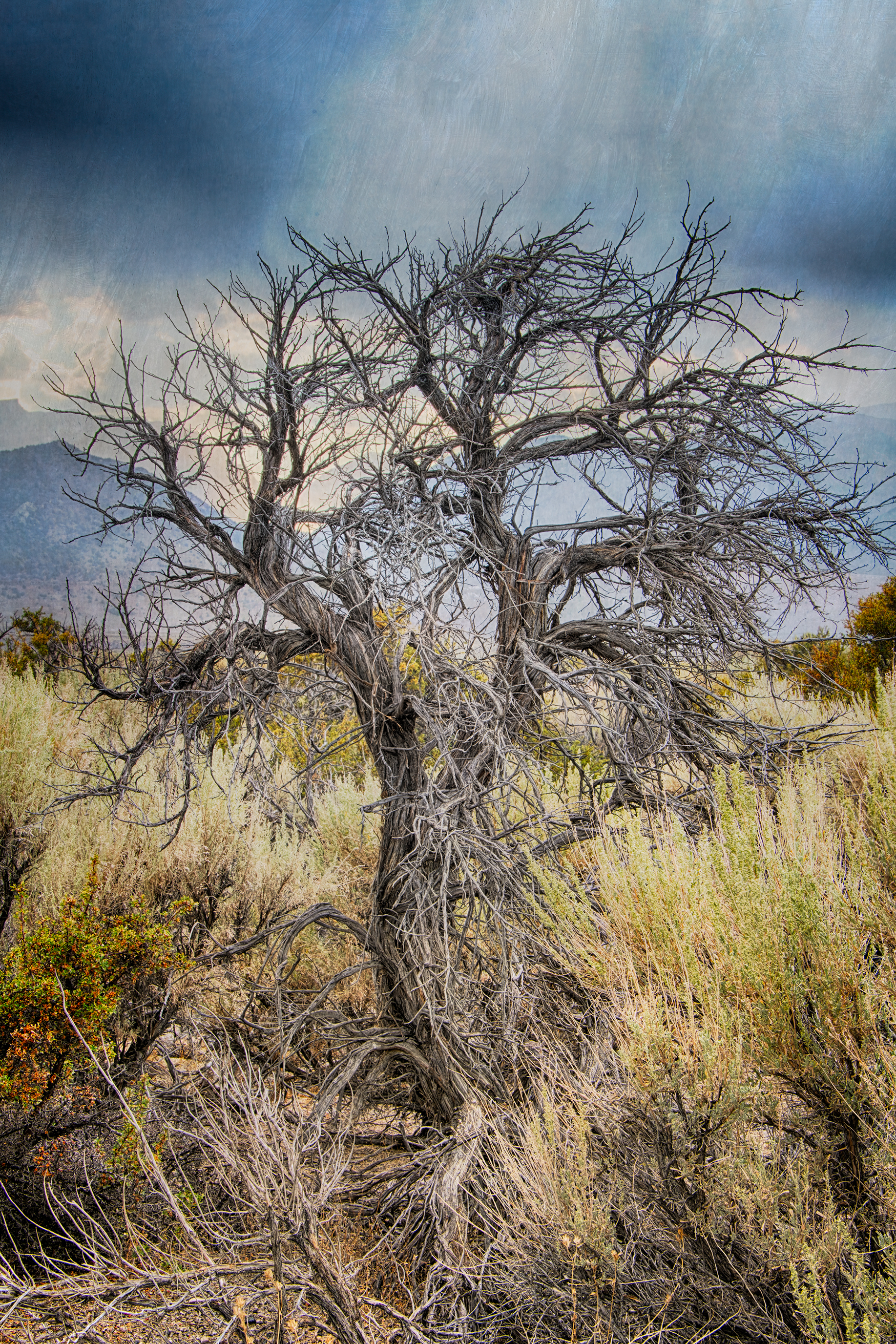
East of the Sierras © Harold Davis


September 3, 2018
Multiple Exposures and Models: Star of Brightness.
The technical idea behind my in-camera series of multiple exposures of models is to work with the models to use the human body to create an external shape, as defined by the models’ positioning when the strobes fire. The philosophic idea is to use external shape creation of the human form to allude to the divine, in a variety of guises and traditions.
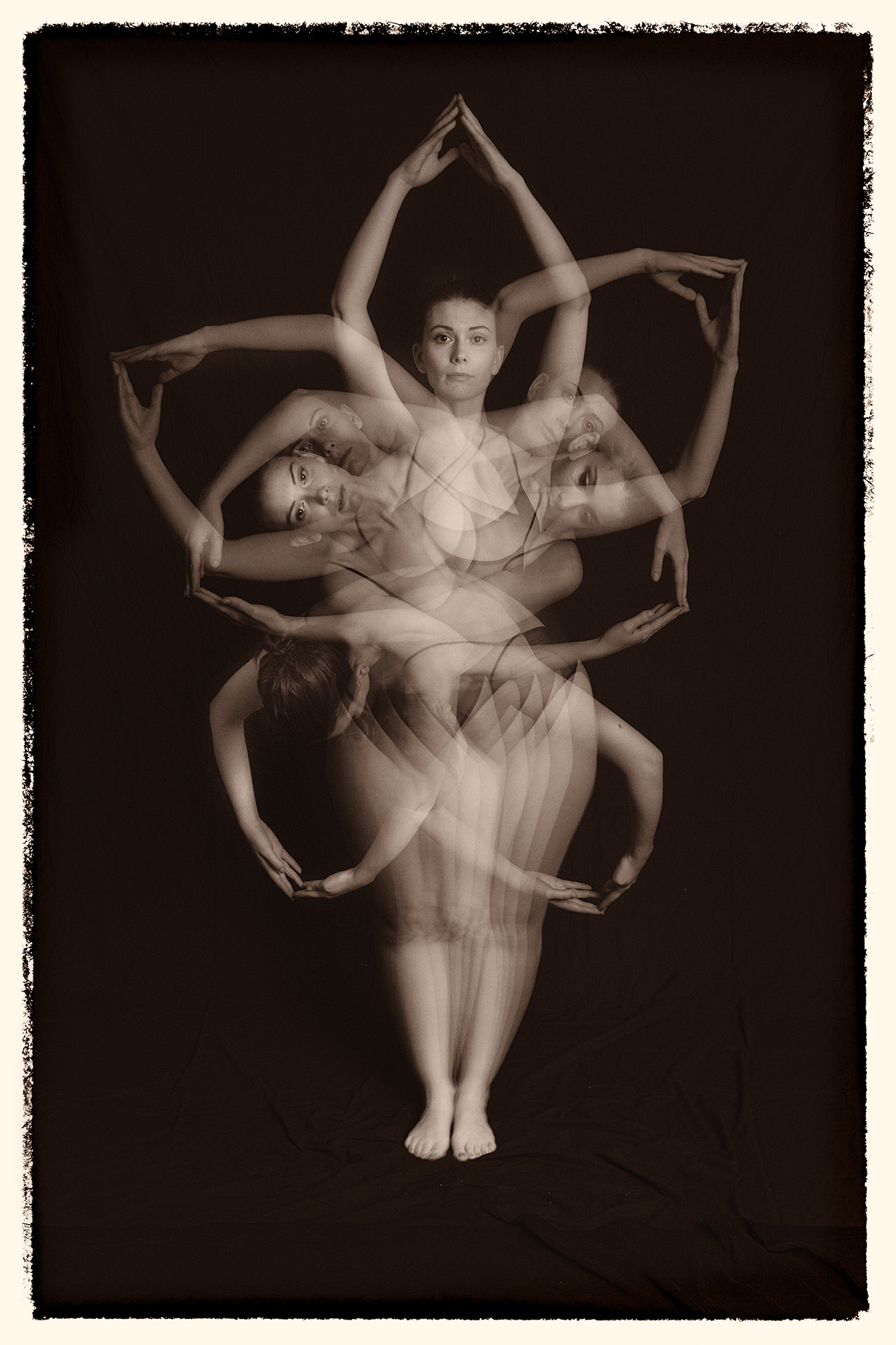
Star of Brightness © Harold Davis
The model in Star of Brightness (above) is Muirina Fae. She’s also the model in Vitruvian Woman, Devotional Pose, and Avatar or Artifact.
The model, and communication with the model, makes a huge impact on this kind of image making. Animism (below) is a collaboration with A Nude Muse, who can also be seen in the White Daemon Series and Once Upon a Tintype.
If you are interested in the archetypes and iconography of this series, check out Introducing Multiple Exposures and the Multiple Exposures online gallery.
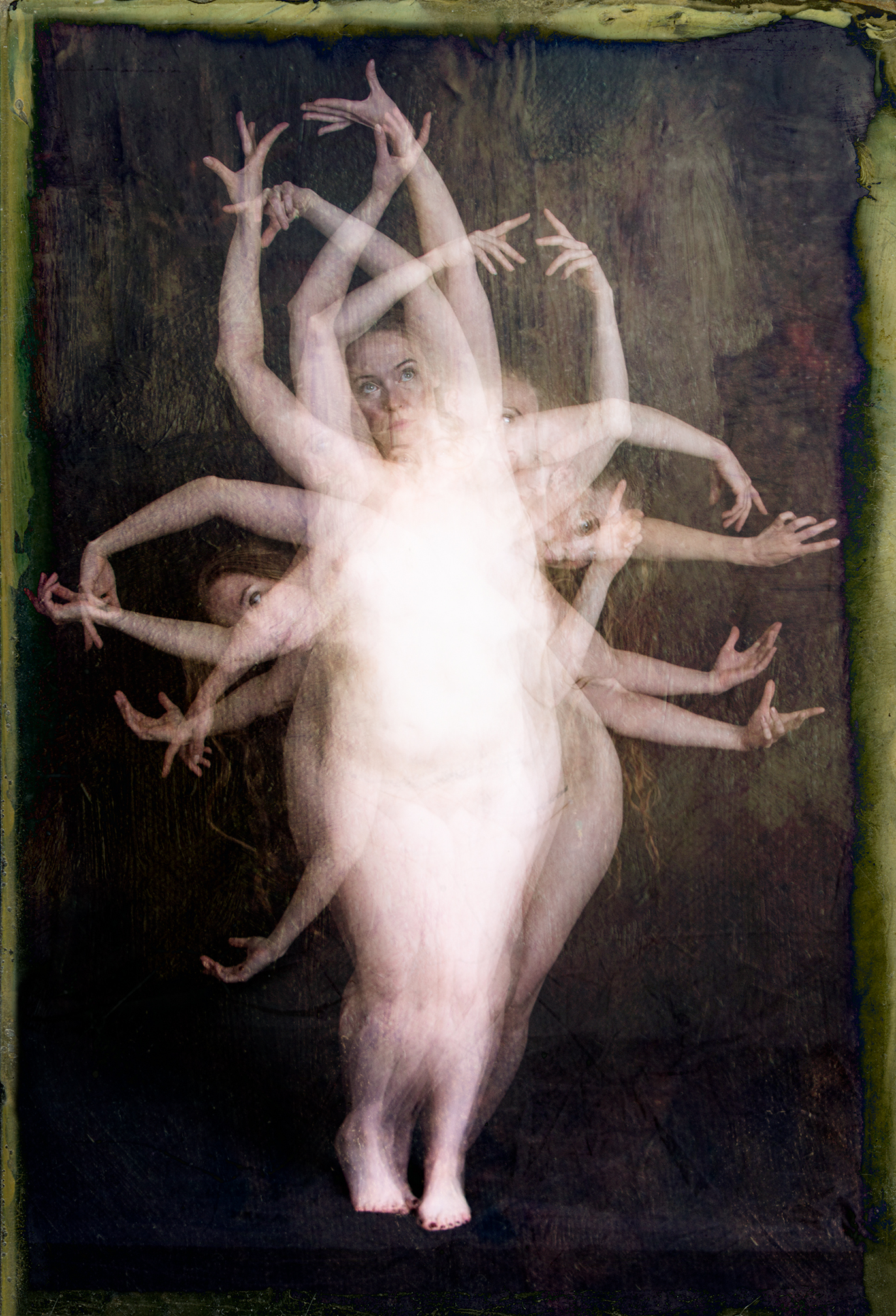
Animism © Harold Davis

September 1, 2018
Frankfurt Station
Like the nearly empty road in Poem of the Road, train stations give rise to existential questions. Where are we hurrying, and why? When a track and platform are empty, has anyone actually traveled that way? When I am travelling by train, I am always wondering about departures and arrivals—if not my own, then the comings and goings of all the other people, who are there, or not there.

Frankfurt Station © Harold Davis
This is an iPhone capture, made while waiting to change trains in Frankfurt, Germany.

August 30, 2018
X-Ray and “Fusion” X-Ray Images of Flowers
Here are some (more) x-rays and fusion x-rays of flowers. Fusion x-rays use post-production to combine “straight” x-rays with light box images of the same composition. For background information on how these images were made, see Revealing the Unseen with X-Ray Photography of Flowers.
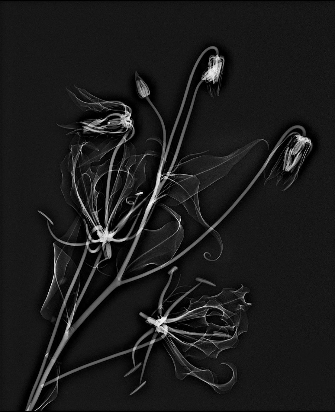
Gloriosa Lily X-Ray © Harold Davis
By way of comparison, here’s a link to one of my digital photos of a Gloriosa lily, photographed on a mirror, with a little background about the flower.
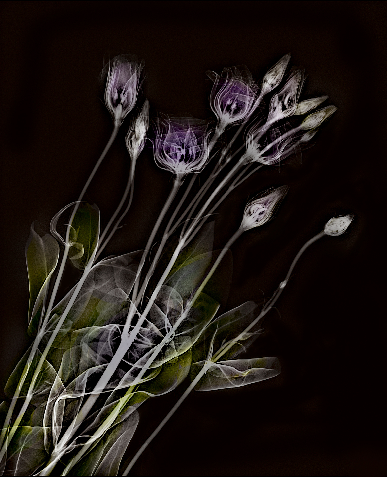
Lisianthus Fusion X-Ray © Harold Davis
These two versions of a bouquet of Lisianthus (above and below) are both fusion imagery, with x-rays and conventional light box photography.
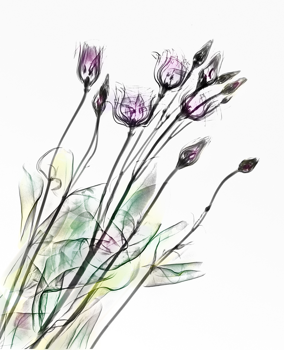
Lisianthus on White © Harold Davis
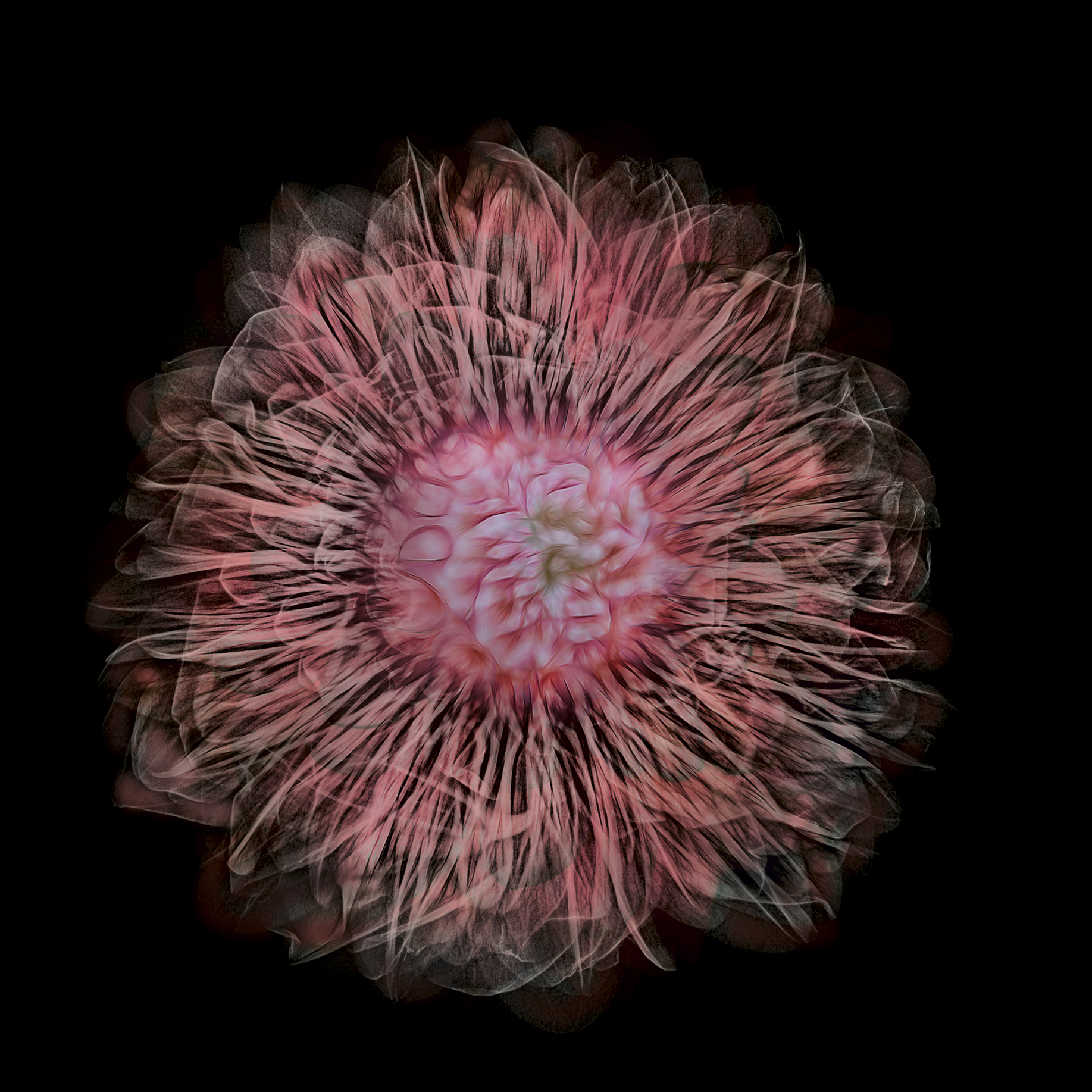
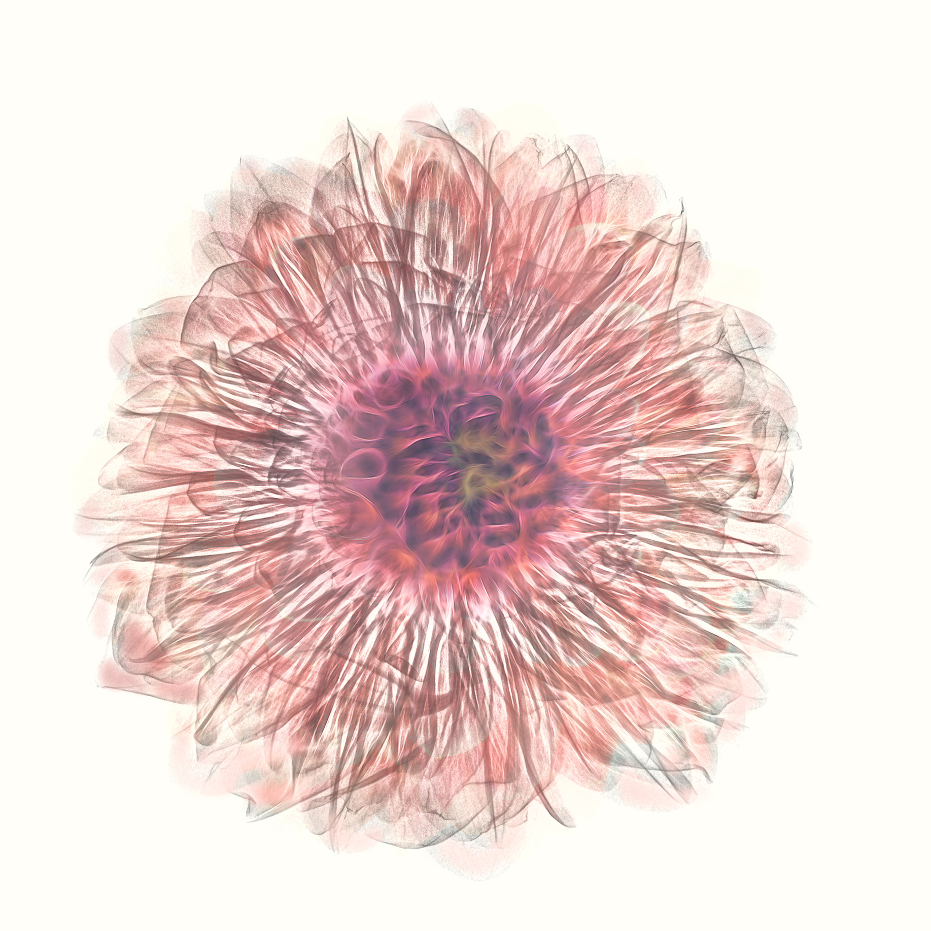
I am pleased that the Dahlia composition (below) does a good job of showing the secrets within the flower, while also capturing the exterior of the flower!
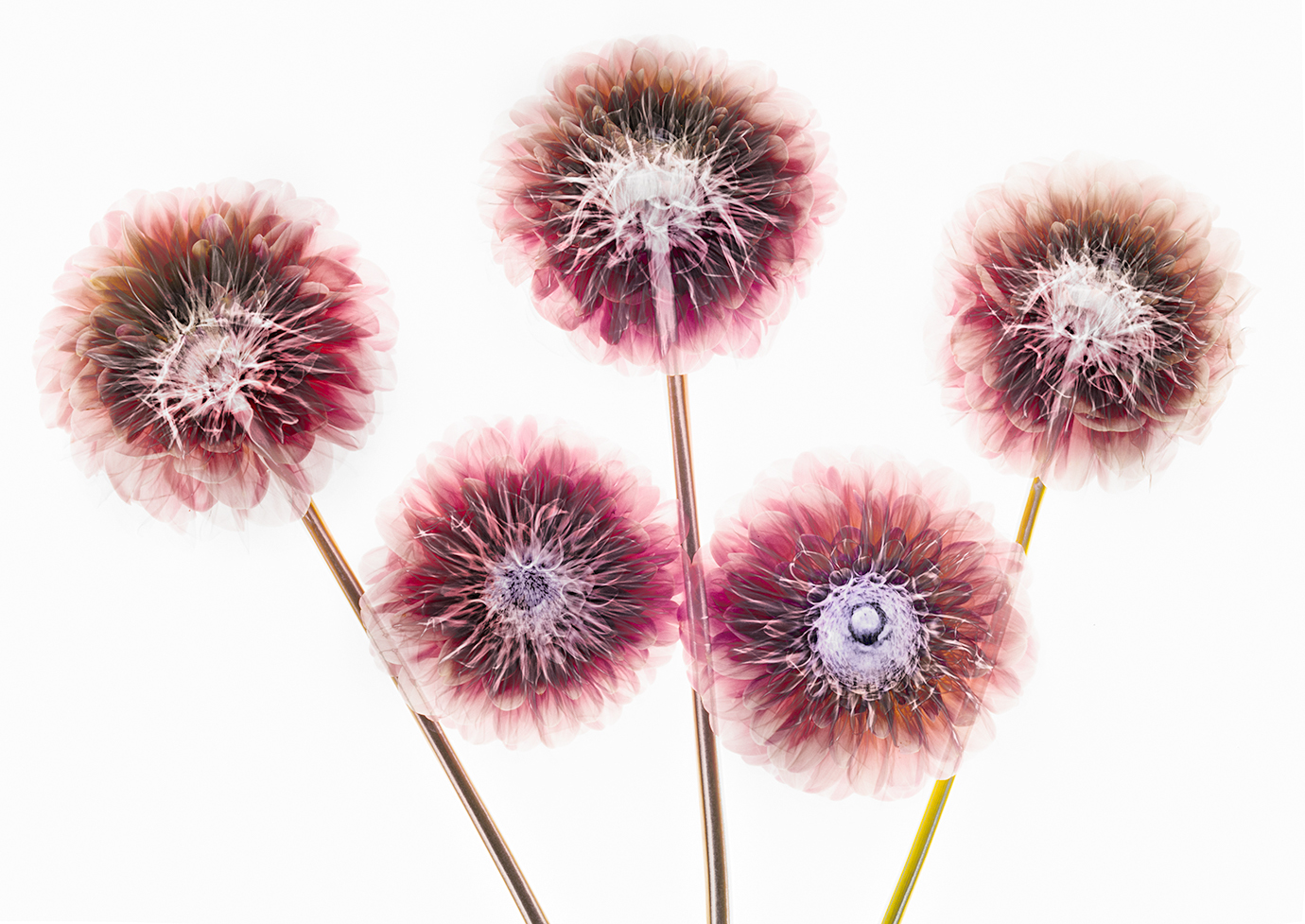
Dahlias Fusion X-Ray © Harold Davis
The sunflower was the first fusion x-ray I processed. I like it also as a straight x-ray (below).
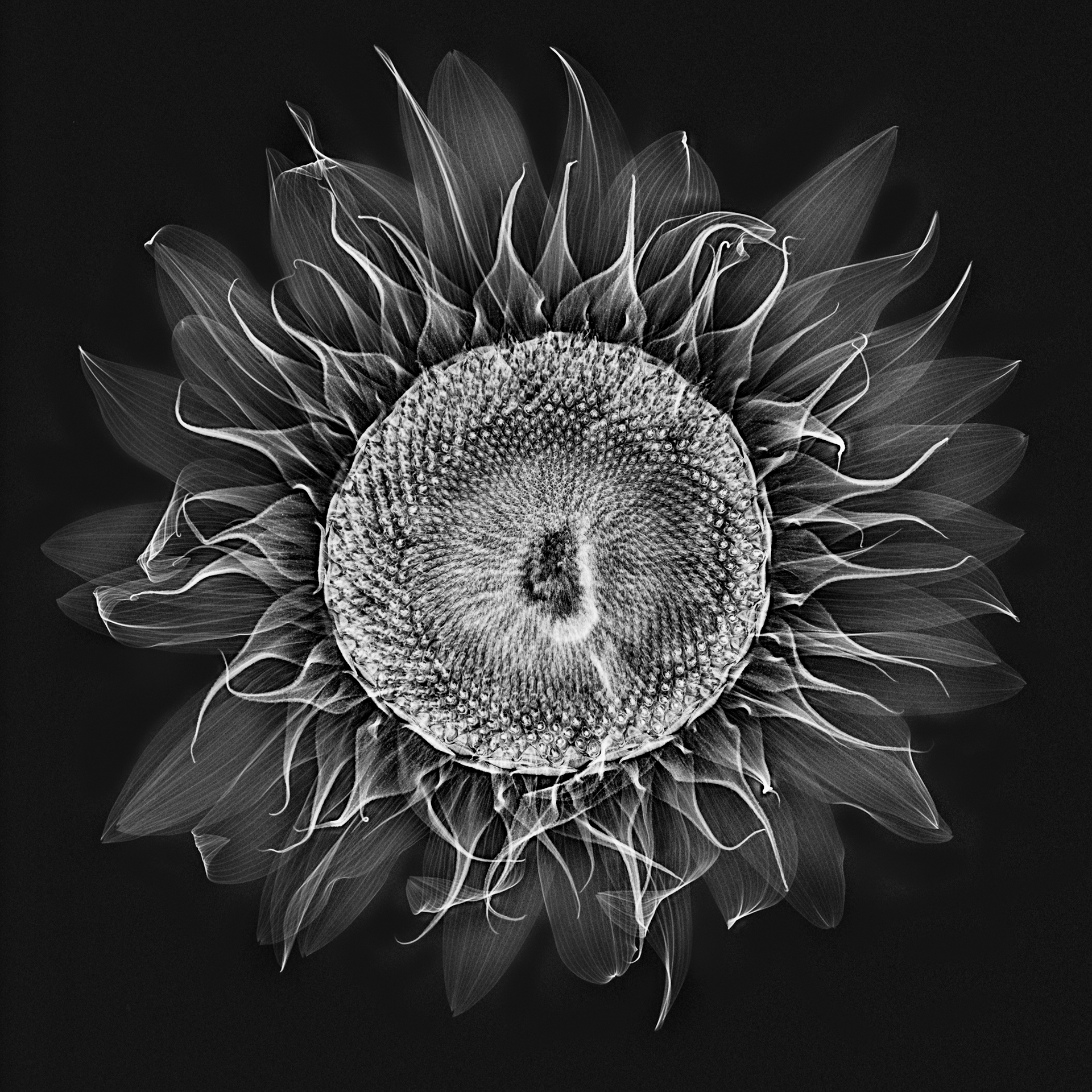
Sunflower X-Ray © Harold Davis

August 28, 2018
Postcards from Berlin
When I got the call that Phyllis had been admitted to the hospital with a mysterious high fever, I knew I had to cut my visit to Berlin and eastern Europe short and come home. My kids didn’t qualify as Boxcar kids, they were Uber kids, journeying via Uber to the supermarket to pick up pasta to cook.
Phyllis is home now and doing so much better (and thanks everyone for the good wishes), the kids were well supported by their cousin and grandparents but were of course very glad to see me. Things are going back to normal.
Anyhow, I had less time in Berlin that I had planned or wanted. Julian (we had been doing x-ray photography together in Heidelberg) was in Berlin with me, and we had a very early morning walk around Berlin’s Tiergarten. The three images in this story are from that walk. Then I took a cab to the airport, flew from Berlin to Munich, and Munich to San Francisco, and hugged the worried kids on the very same day!
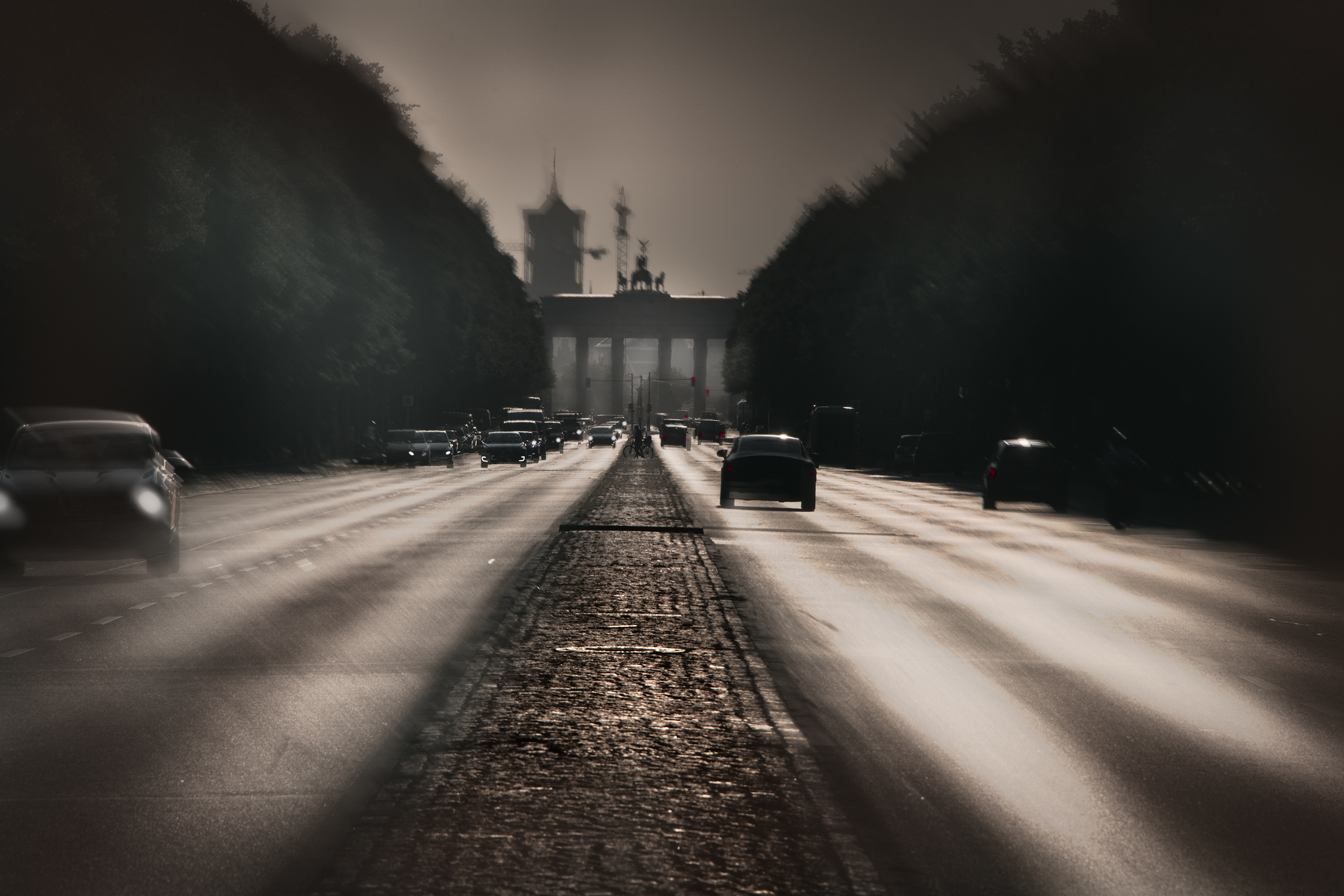
Approaching the Brandenburg Gate © Harold Davis
With the sun coming up from generally behind the Brandenburg Gate, we stopped on a traffic island facing east (photo above). My thought was to process the image to look a little vintage, almost as if it were a relic of the cold war, when the Brandenburg Tor was a symbolic demarcation between east and west Berlin (actually, it was located on the east side of the Berlin Wall).
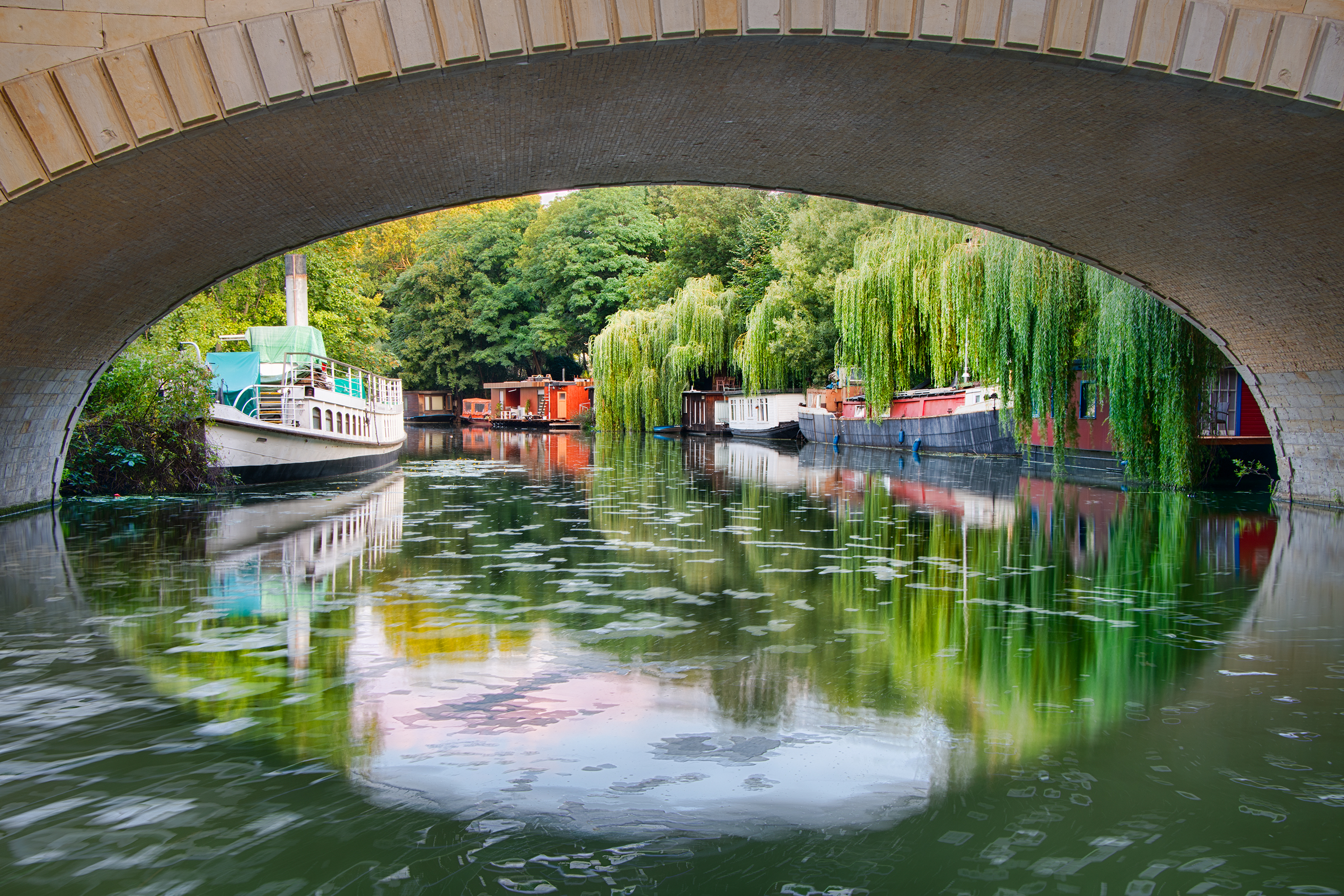
Berlin Canal © Harold Davis
To make this apparently bucolic image of a residential canal framed by the oval of a train bridge (above), we stepped into a small clearing inhabited by sleeping homeless people, who were just starting to rise with the dawn of the new day.

Berlin Reflection © Harold Davis
Across from the ruins of the Kaiser Wilhelm Memorial Church, the lines of architecture are reflected in a city that never seems to sleep, and with much new construction and au courant architecture is an epitome of modernity (photo above).

August 26, 2018
Revealing the Unseen with X-Ray Photography of Flowers
I had to quickly change my plans due to a family emergency and fly home from Berlin. Now that I’m at home, it’s good that things have settled down again, and that I have a chance to review some of the x-ray work I did in Heidelberg in collaboration with Dr. Julian Köpke (you can see some of his x-ray images here).
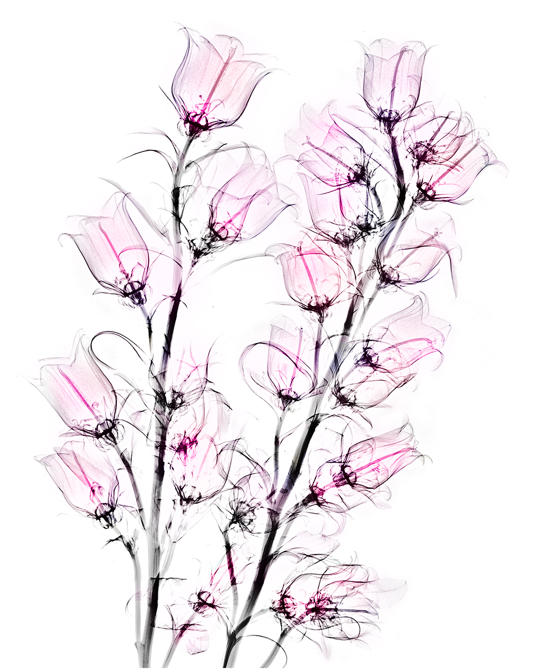
The examples so far on my blog are Campanulas X-Rays, Dahlia Fusion X-Rays and Light Box Photos, More Fusion X-Rays, and Sunflower X-Ray Fusion. Some comments on the process of making these images, what is involved technically, and where we go from here follow the image below (the on-black version of the Campanulas shown here is a good example of a fairly straight x-ray file without additional coloration or fusion with a light box image).
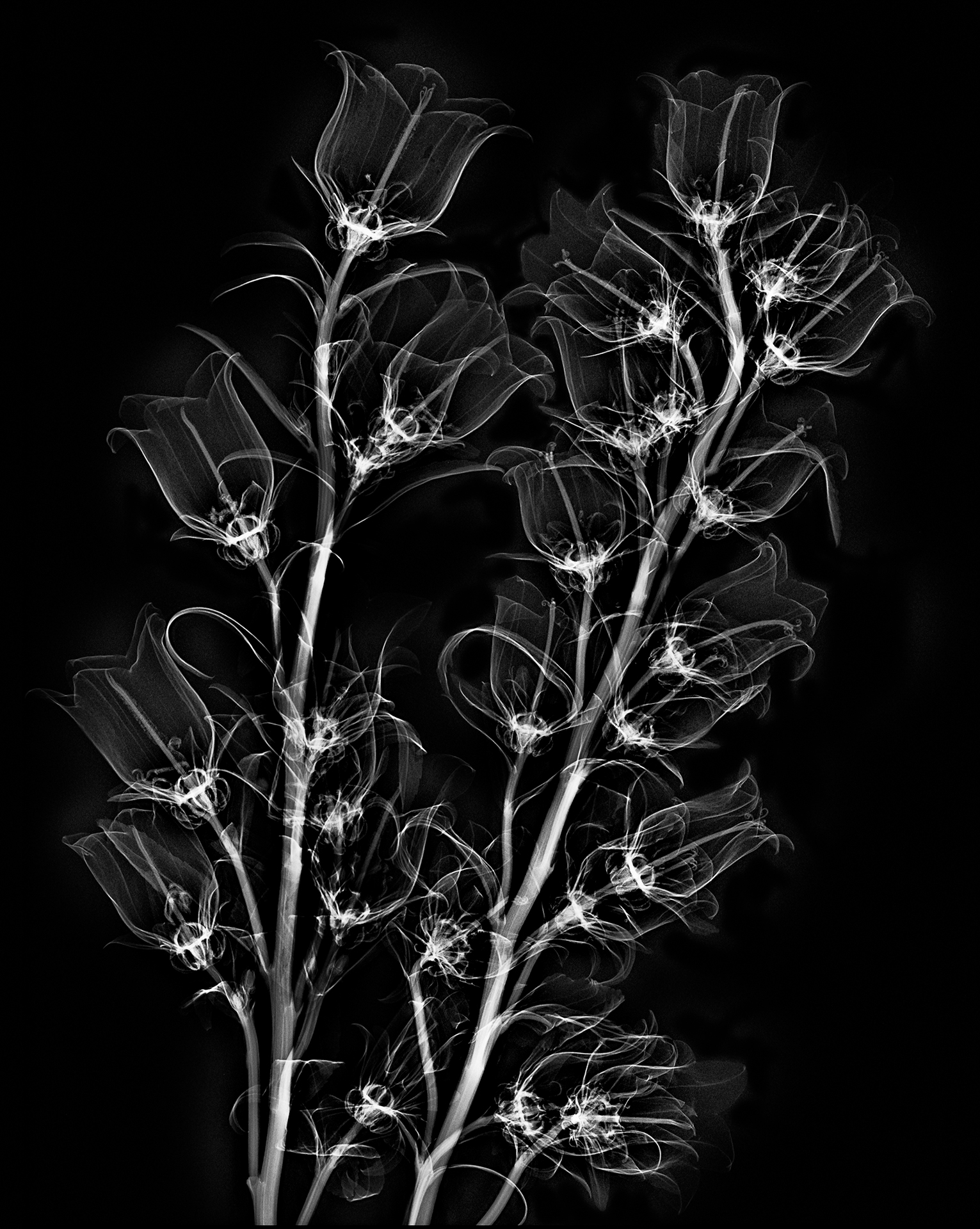
Campanulas X-Rays on Black © Harold Davis
As you likely know, what we see are wavelengths emitted or reflected along the electromagnetic spectrum. Visible light ranges from 400nm to 750nm in terms of wavelength. In comparison, Julian tells me that the x-rays we used had a wave length of roughly 0.04nm (or shorter in wavelength by a factor of about 10,000 than visible light).
Off-the-shelf digital cameras capture roughly the same electromagnetic spectrum as what we see, although it is possible to modify the spectrum that a camera can capture; for example, for Infrared (IR) photography. In addition, even unmodified digital cameras do “see” more of certain frequencies than the human eye, for example, in night photography.
X-rays are a form of electromagnetic radiation not visible to our eyes with a shorter wave length than the visible spectrum. First named and discovered by Wilhelm Conrad Röntgen in 1895 (for which Röntgen won the Nobel prize), X-radiation is primarily used today for medical imaging (also for research and industrial purposes).
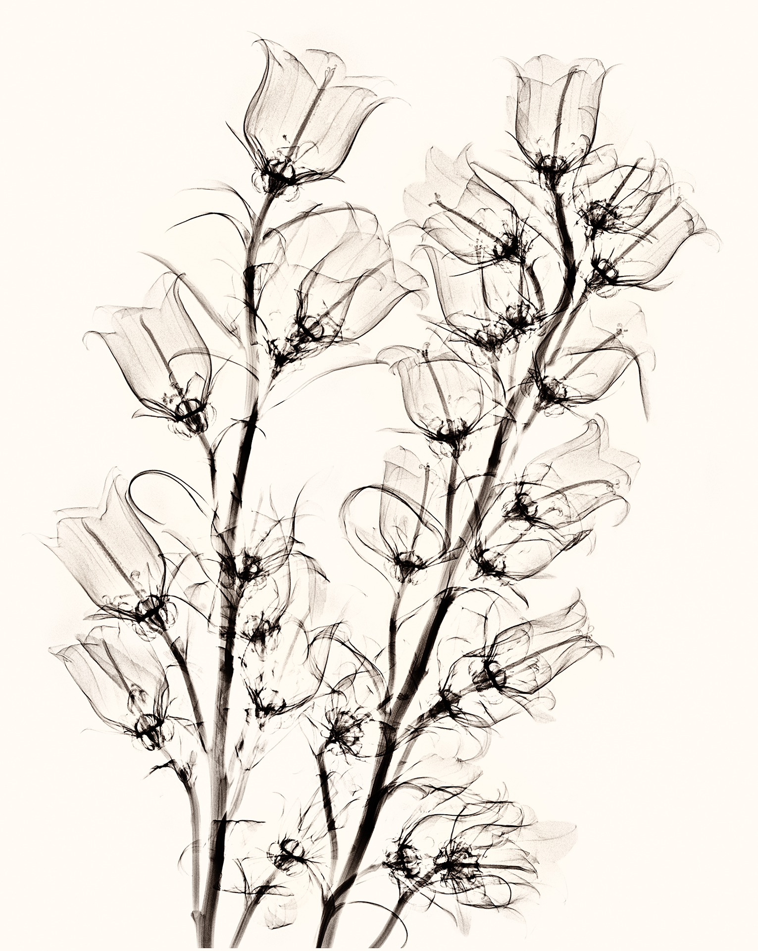
Campanulas X-Ray on White © Harold Davis
Looked at from one viewpoint, X-radiation is just another form of photography using a digital sensor to capture the data generated by exposure to the radiation from an electromagnetic wave. As with any kind of photography (or, indeed, any human endeavor), one of the key ways to get better is to practice a great deal (after all, practicing is how musicians get to Carnegie Hall).
Julian and I had three sessions of x-ray photography of flowers, and by the third session we were beginning to get the hang of the thing.
Our initial idea was to blend x-ray captures of flowers with light box flowers for transparency images. This “fusion” would allow us to show the inside and the outside of the flower composition. We used a clear and rigid plexiglass sheet to align the flowers for both processes.
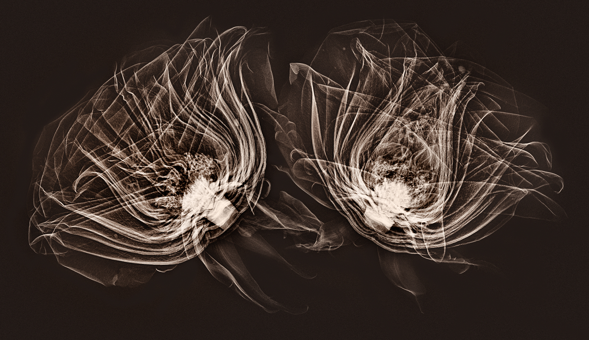
Two Roses (side view) © Harold Davis
The medical x-ray process involves both generating the x-radiation, and capturing it on a digital sensor. In this sense, it is analogous to firing a studio strobe and capturing the light waves emitted on a standard camera sensor. With the stationary medical x-ray device, I was reminded most of an old-fashioned analog darkroom enlarger, where the light beam from the enlarger is captured on media directly below it (photographic paper that is sensitive to light in the analog darkroom process).
We used an x-ray machine designed for mammography for these images.
What turns out is that x-radiation has very different properties as the electromagnetic source than the visible spectrum. X-rays both scatter and decay, in a way that the visible light we are used to does not. So to get good x-ray compositions, it was necessary to work with the characteristics of how the x-radiation emitted from the mammogram system would be captured by the digital sensor, and to arrange the flower compositions accordingly.
To put this comprehensibly, the mammogram system was designed to best capture the shape of the human female breast. We got better results to the extent that our composition could mimic this three-dimensional shape and positioning.
After arranging the flowers on the plexiglass, and aligning the plexiglass with marks we had set up on the machine, Julian and I would huddle behind the operator’s leaded glass shield as Julian operated the machine.
The straight x-ray photos of flowers are really beautiful, and I am grateful to have had the chance to experiment with this. I am also very excited about the fusion imagery, which I have never seen before. I look forward to exploring this area further, and to more collaboration with Julian.
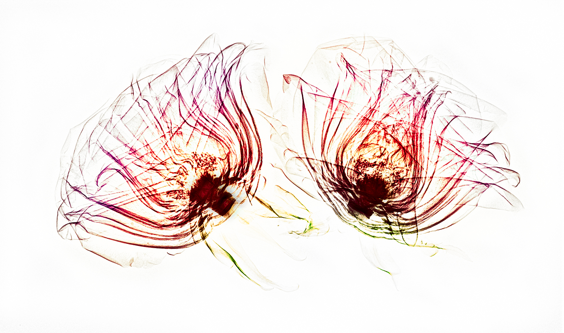
Two Roses (fusion image) © Harold Davis

Revealing the Unseen with X-Ray Photography
I had to quickly change my plans due to a family emergency and fly home from Berlin. Now that I’m at home, it’s good that things have settled down again, and that I have a chance to review some of the x-ray work I did in Heidelberg in collaboration with Dr Julian K. The examples so far on my blog are Campanulas X-Rays, Dahlia Fusion X-Rays and Light Box Photos, More Fusion X-Rays, and Sunflower X-Ray Fusion. Some comments on the process of making these images, what is involved technically, and where we go from here follow the image below (the on-black version of the Campanulas shown here is a good example of a fairly straight x-ray file without additional coloration or fusion with a light box image).
Campanulas X-Rays on Black © Harold Davis
As you likely know, what we see are wavelengths emitted or reflected along the electromagnetic spectrum. This portion of the electromagnetic spectrum, roughly 430–770 THz in terms of frequency, is referred to as the “visible spectrum”.
Off-the-shelf digital cameras capture roughly the same electromagnetic spectrum as what we see, although it is possible to modify the spectrum that a camera can capture; for example, for Infrared (IR) photography. In addition, even unmodified digital cameras do “see” more of certain frequencies than the human eye, for example, in night photography.
X-rays are a form of electromagnetic radiation not visible to our eyes with a shorter wave length than the visible spectrum. First named and discovered by Wilhelm Conrad Röntgen in 1895 (for which Röntgen won the Nobel prize), X-radiation is primarily used today for medical imaging.
Campanulas X-Ray on White © Harold Davis
Looked at from one viewpoint, X-radiation is just another form of photography using a digital sensor to capture the data generated by exposure to the radiation from an electromagnetic wave. As with any kind of photography (or, indeed, any human endeavor), one of the key ways to get better is to practice a great deal (after all, practicing is how musicians get to Carnegie Hall).
Julian K and I had three sessions of x-ray photography of flowers, and by the third session we were beginning to get the hang of the thing.
Our initial idea was to blend x-ray captures of flowers with light box flowers for transparency images. This “fusion” would allow us to show the inside and the outside of the flower composition. We used a clear and rigid plexiglass sheet to align the flowers for both processes.
Two Roses (side view) © Harold Davis
The medical x-ray process involves both generating the x-radiation, and capturing it on a digital sensor. In this sense, it is analogous to firing a studio strobe and capturing the light waves emitted on a standard camera sensor. With the stationary medical x-ray device, I was reminded most of an old-fashioned analog darkroom enlarger, where the light beam from the enlarger is captured on media directly below it (photographic paper that is sensitive to light in the analog darkroom process).
We used an x-ray machine designed for mammography for these images.
What turns out is that x-radiation has very different properties as the electromagnetic source than the visible spectrum. X-rays both scatter and decay, in a way that the visible light we are used to does not. So to get good x-ray compositions, it was necessary to work with the characteristics of how the x-radiation emitted from the mammogram machine would be captured by the digital sensor, and to arrange the flower compositions accordingly.
To put this comprehensibly, the machine was designed to best capture the shape of the human female breast. We got better results to the extent that our composition could mimic this three-dimensional shape and positioning.
After arranging the flowers on the plexiglass, and aligning the plexiglass with marks we had set up on the machine, Julian and I would huddle behind the operator’s leaded glass shield as Julian operated the machine.
The straight x-ray photos of flowers are really beautiful, and I am grateful to have had the chance to experiment with this. I am also very excited about the fusion imagery, which I have never seen before. I look forward to exploring this area further, and to more collaboration with Julian K.
Two Roses (fusion image) © Harold Davis
August 22, 2018
Campanulas X-Rays
I write this from the train from Frankfurt to Berlin, where Julian K. and I are sitting in a first class car at the all-important table, with the all-important wi-fi connectivity. Since I am connected to the world of the Internet, I can post this!
Campanulas X-Ray © Harold Davis
We are working on processing images from yesterday’s fusion light box and x-ray photography of flowers. These campanulas as arranged took up almost all the space yielded by the size of the sensor of the x-ray machine. I’ve begun to get a bit of a feeling for how the x-ray sensor processes the electromagnetic waves it receives from the emitter, and how to best arrange the subject in the cone shape that is best for this kind of capture.
Query: Is capture via x-ray photography, or should another term be used? I think “photography” does fit the bill (there is another, more technical vocabulary related to radiology as well). What do you think?

August 20, 2018
Dahlia Fusion X-Rays and Light Box Photos
Dahlia Fusion X-Ray Inversion © Harold Davis
Dahlia Fusion X-Ray © Harold Davis
This Dahlia was photographed on a light box for transparency, then captured via x-ray photography. The two capture techniques were combined in Photoshop. In the upper version, there is also an L-channel inversion in LAB color.
I worked with my friend Julian Kopke, who is a medical doctor, radiologist, and a physicist to create these images.
This technique is also shown here, and with a Sunflower.

August 17, 2018
Top Photography Blog Honor; More Fusion X-Rays
I am honored to be included as one of the top fifteen photography blogs in the English language worldwide. This is good company to be in. Thanks for the award!
Moving on, here are two more fusion light box (Flowers for Transparency) and x-ray images, one of Cala Lilies and the other of Alstroemerias. It has been really fun working with Julian to create these images, and I look forward to using conventional cameras and medical x-ray imaging gear to make and process more imagery.
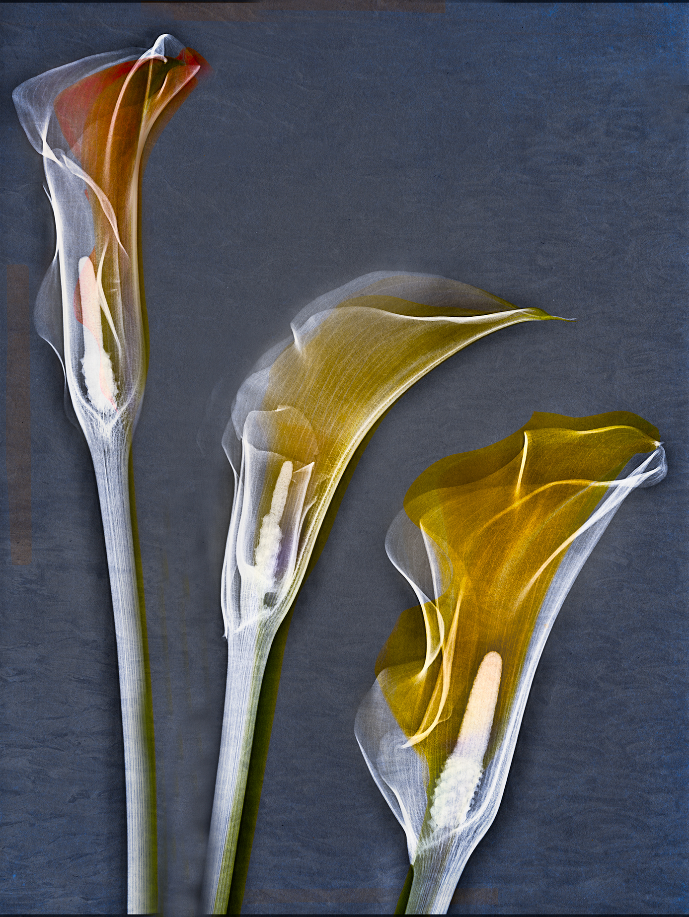
Cala Lilies Fusion X-Ray © Harold Davis
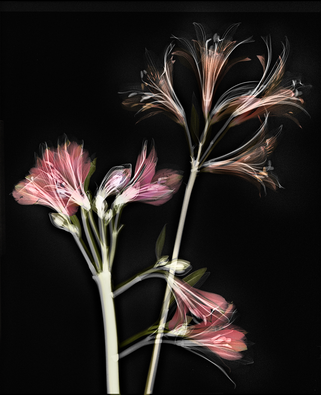
Alstroemerias X-Ray Fusion © Harold Davis


